(1)
Swingle Clinic, Vancouver, BC, Canada
Clinical Versus Normative Databases
For clinicians, the most accurate databases are clearly clinical. Normative databases are far less accurate. The fundamental organizing concept of the normative database for the clinical practitioner is, simply stated, wrong.
The organizing concept for normative databases is that one can identify a group of individuals who are symptom free and therefore have “normal” functioning neurology. This group of symptom free individuals then serves as the comparative database to identify those who are statistically discriminant. The statistical departures from the normative database define the anomalous neurological condition that is associated either causatively or exacerbatitively with the client’s clinical condition. This concept is wrong.
The reason that normative database treatment recommendations are so often incorrect is because the fundamental premise is wrong. Symptom free individuals may well have predispositions to conditions that have not manifested. The data are quite clear and we have definitive evidence for this that spans decades.
Let us simply take the example of heritability data for schizophrenia. As the data in Table 2.1 indicate, if one monozygotic twin has been diagnosed with schizophrenia the probability that the second identical twin will have schizophrenia is about 50 %. But, the interesting statistic is that 50 % will not! Where do we find the 50 % without manifested schizophrenia, but obviously with the same genetic load? In the normative databases! So clearly the organizing concept for normative databases, at least for clinicians, is incorrect. Normative databases so constituted ignore basic psychopathology and basic biology. Every person has predispositions. Predispositions to anxiety, depression, emotional volatility, and the like. However, many of these predispositions are not manifest at any particular time. In general, clinicians understand that one needs an experiential trigger to “turn-the-key” to manifest a neurological predisposition.
Table 2.1
Heritability statistics on schizophrenia
Genetic predispositions | |
|---|---|
Monozygotic twins | 30–50 % |
Dizygotic twins | 15 % |
Siblings | 15 % |
General population | 1 % |
Adopted-biological relatives with Schizophrenia | |
|---|---|
Adoptee with Schizophrenia | 13 % |
Adoptee without Schizophrenia | 2 % |
These logic considerations are well known and surprisingly, at least to me, ignored by non-clinicians that develop the normative databases. If in the normative database one has subjects with non-manifested predispositions, then statistically one can expect very poor discrimination.
Conditional Probability Models
There are many conditional probability models associated with the concepts of differential susceptibility. In mathematical game theory, the probability of future actions is predicated on present state. In chess, the probability of Queen move is markedly different if Queen Pawn has advanced. This is considered a state conditional probability.
In optimal performance contexts, conditional probability theories consider both vulnerability as well as resilience markers. The markers can be direct, or primary, such as the genetic serotonergic system inefficiency affecting stress tolerance. The concept of “preparation for duty” for military and police personnel is premised on reducing vulnerability to work stress (e.g., combat) by increasing the neurological basis for stress tolerance.
Secondary markers may be introversion that reduces probability of development of social relationships that in turn is negatively synergic with the primary marker. Hence, in the latter case the individual who has experienced severe stress may be more vulnerable to negative posttraumatic sequellae if the secondary marker impeded the development of a social support network.
Obviously, in the clinical context, individuals who present themselves for treatment have a manifested susceptibility factor. Individuals who do not present for treatment may have the same neurological predisposition but has not manifested. Hence, the latter individual is a candidate for normative database whereas his cohort with the identical, but manifested, predisposition is in my office and hence in the clinical database. Also, obvious, the normative database is going to be statistically blind to many neurological conditions that are predispositions.
Where normative databases have strength are determinant neurological abnormalities such as those associated with epilepsy, autism, structural damage, and progressive neurological deterioration. Conditions associated with primary genetic (e.g., dopamine/serotonin), secondary endophenotypic (e.g., autonomic reactivity) and phenotypic (e.g., sensory processing), and tertiary endomorphic (e.g., body mass) are likely to be under the statistical discrimination thresholds. However, most importantly, the normative databases just simply miss neurological relationships found in brainwave activity for conditions that bring clients into the clinician’s office.
The ClinicalQ is a clinical database. The database contains 1,508 clinical clients. The organizing logic is that clients who report a condition (e.g., depression) have a neurological representation of that condition. Based on the diathesis vulnerability model, the condition reported by the client is one that is associated with a neurological predisposition that has manifested. A normative database is likely to miss this entirely since this clinical client, before becoming depressed, had the same neurological predisposition but would be considered “normal” (i.e., symptom free) and eligible for the normative database.
The important concepts of the vulnerability or conditional probability models for the clinician include conditional vulnerability (cf., Ingram and Luxton 2005), diathesis (Sigelman and Rider 2009; Belsky and Pluess 2009) and that although neurological predispositions are stable across the lifespan, they are not unchangeable (Lipton 2006; Oatley et al. 2006).
Although the theoretical concepts associated with predispositions and vulnerabilities are of interest, for the purposes of this guide, the critical issues are that predispositions are just that, predispositions. It is also important that predisposition does not mean inevitable. People can have a multitude of predispositions but may be fortunate enough to never have them triggered and therefore be even more fortunate to never need our services.
Finally, expressivity of the predisposition in neurology is analogous to severity of a condition in clinical medicine. The severity of the EEG condition is not directly associated with the severity of the symptom. In general, the more severe the EEG condition, the more pronounced the symptomatology in terms of several parameters including chronicity, intensity, treatment resistance, and qualitative manifestation. However, many variations occur so that clinically one uses the ClinicalQ to identify clinical conditions that should be probed/explored with the client. The qualitative features of the symptoms may well be poorly correlated with the magnitude of the ClinicalQ markers. This is especially true of ClinicalQ markers associated with experiential factors as compared to genetic predispositions.
The ClinicalQ
Introduction
To illustrate the superiority of clinical norms, consider the following comparison with a normative database (Figs. 2.1, 2.2, and 2.3). Both the ClinicalQ and the 19-point full EEG were obtained simultaneously. The normative report was generated by one of the best known services whereas the ClinicalQ was generated immediately while the client was still hooked up. Many manufacturers of EEG platforms have software available for generating the ClinicalQ data and probes; however, following the outline in the Appendix one can generate the ClinicalQ data and summary with any EEG platform with the aid of a desktop hand calculator.
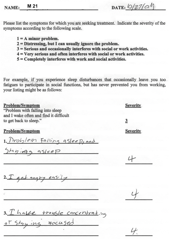
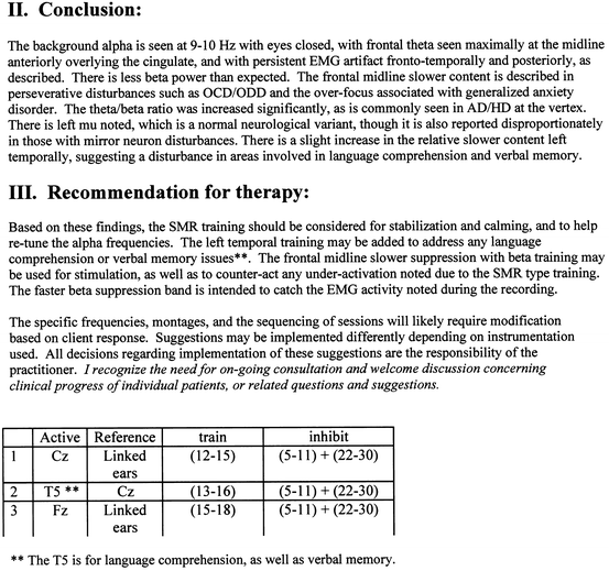
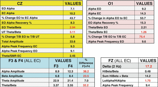

Fig. 2.1
Client’s (M21) self-reported conditions

Fig. 2.2
Client M21. Full 19-site QEEG report from independent service using normative database

Fig. 2.3
ClinicalQ for client M21
It is quite apparent that the ClinicalQ was far more accurate for this client. He reported sleep problems consistent with the low Theta/Beta ratio under eyes-closed conditions at location O1. The marked imbalance in Alpha, frontally, with Alpha being considerably higher in amplitude in the right relative to the left is the marker for emotional volatility. As this client reports: “I get angry easily.” The client’s complaints of problems with focus and attention are reflected in the elevated Theta/Beta ratios at location Cz, F3, and F4 as well as the elevated Delta and slow Alpha as measured at Fz.
We also see another marker that is not reported by the client. Beta is considerably greater in amplitude in the right relative to the left frontal cortex. This is a marker for depression. When probed about this, the client admitted to feeling “low” much more intensely and frequently then he believed was the case with his friends.
The ClinicalQ shows precisely where to treat these conditions and what to treat: Standard Theta/Beta training at locations Cz, and if necessary later at F3 and F4. Increasing the Theta/Beta ratio at O1, eyes closed, for the sleep problems. Speed up the Alpha peak frequency (or decrease the amplitude of low Alpha) and finally balance the frontal regions, F3 and F4, in the Alpha and Beta ranges. Rule of thumb—treat sleep problems first as restored sleep quality is likely to result in other improvements in brain functioning. There are many other general guidelines for how to approach developing a treatment strategy for the clients that will be discussed more specifically later in this book.
It is also apparent that the QEEG report not only did not identify the client’s complaints but the treatment strategy recommended is largely irrelevant to the client’s problems. The possible exception is the recommended 12–15 Hz training at Cz. However, neurofeedback at almost any location is usually associated with client reports of improvement early in treatment.
So, it is obvious that the ClinicalQ is not a poor practitioner’s substitution for the full 19-site QEEG. Many mini-Q systems are being marketed on exactly that basis. The purpose of using the ClinicalQ is to make neurotherapy much more efficient; because, again, the ClinicalQ is more accurate for clinical practice than the normative databases.
The intake procedure with the ClinicalQ is the first therapy session. Clients are strongly relieved that their complaints are understood, that there are identifiable neurological causes/corollaries of their condition, and there is a precise “game plan” for treatment. The average length of treatment at the Swingle Clinic for most conditions is about 23 sessions, and for simple problems like Common ADD (CADD) it is closer to 15, which is far below industry standards (Swingle 2002). This efficiency is based on many aspects and modalities of treatment such as the use of braindriving techniques and the home treatment procedures described in later chapters. However, a major contributor to that efficiency is the ClinicalQ at the initial visit.
When clients present for treatment, they generally expect to have to spend at least one, perhaps several, sessions telling their tales of woe and submitting to various forms of assessment. Imagine their pleasant surprise when they experience something so radically different, yet so logically sound, as letting the brain diagnose their problem. As I will detail later in this chapter, one can literally tell the client why he or she came to your office without any report from them and with less than 10 min of EEG recording.
Again, the reason for the unique accuracy and efficiency of the ClinicalQ is because this assessment is based on clinical norms. For all of the reasons discussed above, for intake of clinical clients, normative databases are simply too imprecise.
After telling clients why they are sitting across from you, you can point out how you knew what the problems were and exactly what areas of brain functioning are going to be modified to correct those problems. Treatment can start immediately, certainly in terms of home treatments, and often with brief neurotherapy at the very same first visit. Quite a departure from their experience with other healthcare providers. In addition, the client knows that brainwave assessment has efficacy because you have accurately and completely described the symptoms relying only on the ClinicalQ.
A number of clinicians have shown that there are identifiable EEG patterns associated with a variety of physical and psychological disorders. Deficient Alpha power in schizophrenics has consistently been reported, for example, and EEG slowing is a good indicator of degree of cognitive impairment (Hughes and John 1999). Similarly, specific EEG patterns have been shown to be associated with various forms of ADD (Thompson and Thompson 2006; Swingle 2010), learning disabilities (Thornton 2006), and physical disorders (Hammond 2006a). Thus, like neurofeedback, diagnostic use of the EEG has a research base extending over many years.
The database for the ClinicalQ diagnostic procedures is very large in comparison to some of the previous studies. With over 1,500 patients, the sample size for the ClinicalQ database exceeds even some of the QEEG databases. Further, the ClinicalQ procedure avoids diagnostic labels and categorizing but focuses rather on the behavioral manifestations of the inefficiencies found in brain activity.
When clients present for treatment, the vast majority do not really care if their brainwave architecture departs from normative values. Moreover, sometimes the problem resides in brainwave activity that is not outside normative ranges as determined by the databases. For the reasons discussed above, normative ranges may be statistically insensitive to discriminative patters associated with symptoms. Hence, reliance on normative databases can result in missing areas of opportunity for neurotherapy.
Clients want their problems resolved regardless of normative EEG values. Many clients have endured various healthcare providers’ efforts to deal with their problems, often with repetitive intake evaluations that are time- and money consuming. Imagine how they might feel about your potential for helping them if you tell them more about their problems within 30 min than others have been able to tell them after many sessions and/or assessment procedures.
Dramatic instances of clients being shocked by this highly efficient intake procedure occur when the ClinicalQ record shows the “trauma signature.” The details associated with the trauma signature will be described in detail later in this chapter, but basically it is a marked absence of Alpha amplitude under eyes-closed conditions. Imagine the client’s surprise when, after a few minutes of recording, the practitioner asks if they have a history of emotional trauma. The accuracy rate associated with the trauma signature is good. In the words of one physician who recently introduced the ClinicalQ into his practice, the procedure vastly expands the therapeutic options because of a profoundly expanded understanding of the patient, which the patient recognizes.
Words from a Mom on the ClinicalQ Assessment
Susan Olding
From her book “Pathologies” Freehand Press
Desperate, determined, undeterred by cost or lack of insurance coverage, undismayed by the doubts of conventional physicians, undaunted by the practitioner’s Dickensian-sounding name, I switched off my cell phone at the threshold of Dr. Swingle’s office and carried my daughter across…


I had brought a medical and developmental history—the long litany of concerns that had brought us to his door—but Dr. Swingle waved the papers aside without even looking at them. Instead, he ushered Maia toward a computer screen on the other side of the room and told her to put her feet on the stool below. Then he fixed a couple of delicate wires to her ears…
Then Dr. Swingle sent Maia to the “treasure chest” in the waiting room. He stared at the printout in his hand. “Here,” he said, and he pointed to an outline of the brain, “These numbers imply trauma.” He shrugged, palms up, waiting for my response. I nodded. “And here,” he continued, “too much theta. This is the hyperactivity people associate with ADHD. But it’s minor. In the ballpark I play in, she barely makes the field.” There was more: extreme stubbornness, a tendency to perseverate, lapses of short-term memory, attachment disorder, inability to read social cues, emotional reactivity, tantrums, explosions. One by one he read the ratios, divining1 my daughter’s character—more quickly, more accurately than any professional I’d yet encountered.
The ClinicalQ does not replace the full QEEG. We often do a full QEEG on clients after 10 or so sessions. We do so to assess therapeutic progress but also to provide further interpretative opportunities offered by 19 sites of data. For example, many clients have difficulties that may be more efficiently addressed by treating problems with coherence in the brain. Coherence refers to the degree of interaction, or communication, between brain sites. Hyper-coherence is when the brain sites are not functioning in an efficient interdependent fashion, but rather have too much “cross-talk.” This condition is often found with brain injury, after which clients experience stereotypical, perseverative, and inflexible behavior and cognitive processing. Hypo-coherence, poor inter-site interaction, is associated with diminished cognitive efficiency. To assess coherence in the brain, a full QEEG is required.
However, even if I start with a full QEEG, I always provide the client with immediate feedback based on the data analyses of the ClinicalQ. I do so, as stressed above, for purely therapeutic reasons. Even though clients may have to wait for the QEEG assessment, as they do for virtually all other medical and psychological tests, they get immediate feedback regarding their major complaints on the spot with all of the benefits discussed above. Further, more than 80 % of our clients never need the full QEEG because of the efficiency of the ClinicalQ.
The ClinicalQ Procedure
As with any assessment, it is very important to follow the procedure precisely. It is critical to the interpretative probes that the brainwave ranges and EEG sites are as specified in this guide. Unfortunately, some EEG systems have fixed ranges that are slightly different from these and are very difficult to modify. Any deviation from the specified brainwave ranges and EEG sites reduces the efficacy of the procedure.
The length of each measurement epoch is usually 15 s, but this can be modified if necessary. For example, if assessing a child who cannot stay still, one can shorten the measurement epoch and select those with minimal movement.
All of the measurements are in microvolt amplitude, and the ratios are robust across different systems. The summated values, such as Total Amplitude (TA) (i.e., sum of three brainwave bands as described below), may vary from system to system, and certainly among the different filtering options, so the clinician may find it necessary to find equivalent power ranges.
The essential brainwave ranges to measure are Delta (2 Hz), Theta (3–7 HZ), Alpha (8–12 Hz), Sensory Motor Rhythm (SMR) (13–15 Hz), Beta (16–25 Hz), HiBeta-Gamma (28–40 Hz), Lo-Alpha (8–9 Hz), and Hi-Alpha (11–12 Hz). All these ranges need not be measured at all sites. The ClinicalQ only requires three bands at any particular site. The sites are Cz, O1, F3, F4, and Fz, using the 10–20 international system, as shown in Fig. 2.4.
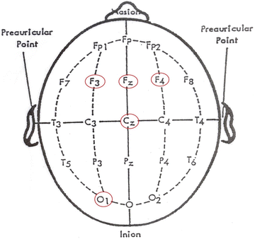

Fig. 2.4
10–20 international EEG site location system. The five-point ClinicalQ locations are noted in red (Colour figure online)
To illustrate the bottom-up assessment procedure, as described by Susan Olding in the above excerpt from her book, consider the following data from an actual client. As emphasized by Susan Olding when I assessed her child, I know nothing about this child other than he is 14 years old and not at all happy about having been dragged into my office by his mother. Let’s call him Mitch, not his actual name, of course.
As noted in the schematic shown in Fig. 2.4, these numbers are from five brain locations: the top of the head (Cz), the back of the head (O1), and the left (F3) and the right (F4) and the middle (Fz) of the front of the head. The measurement requires just over 6 min of recording time and the tasks are simple (open and close the eyes and read something out loud).
In our workshops teaching other clinicians to use the clinical QEEG procedure, we always emphasize that neurotherapy is not a standalone procedure. In short, I tell clinicians “Don’t leave your clinical hat at the door when you do neurotherapy.” During the first session with this child, for example, all I had to do was look at him to know that he was experiencing difficulties. He was sullen and would not make eye contact with me. He slouched in the chair, looking as disinterested as he could, looking out of the window and yawning. His mother was anxious to tell me all about his difficulties.
It is very important in these circumstances not to validate the child’s expectations. What this child was expecting was for his mother to go through her tale of woe telling me all of the difficulties that the child has had and all of the difficulties she and/or the family has had with regard to the behavior of this child. As described in Susan Olding’s account of her experience with the ClinicalQ, I did not permit the mother to proceed with her account describing the child’s behavior; rather, I addressed the child directly.
Often with young children, if you address the issue of sports, you can start to develop some sort of relationship. Unfortunately, in our present digital culture this is becoming less likely as many children, particularly those we see for treatment, are addicted to the internet and have little interest in sports. In this case, I asked the child what sports he played and I was very fortunate that he mentioned soccer. This provided me with my first possible inroad to being able to get this child to acknowledge his difficulties and address the problems. I pointed out that the team that had won the World Cup in soccer in the year 2006 (soccer team from Milan, Italy), every player on the team had done neurotherapy—the same therapy with which he is likely to be involved. I went on to describe some of the other uses of neurofeedback including the local hockey team and the Olympic athletes who were going to participate in the Winter Olympics in Vancouver, Canada. At this point, the child was attentive to me but still rather solemn and not responding with anything but grunts and head nods.
After this brief introduction, I brought the child over to the area where we do the brain assessment and simply told him that he would feel nothing, that this was measurement only, and that it would not take much time. I also pointed out that I would be asking him to open and close his eyes at various times and to read something out loud. I also pointed out that the measurement is movement-sensitive and to try to be as still as possible during the measurement procedure.
The raw data shown in Fig. 2.5 are the result of that assessment. The raw data consists of 99 numbers, and these 99 numbers are reduced to 30 summary markers that are shown in Fig. 2.6.
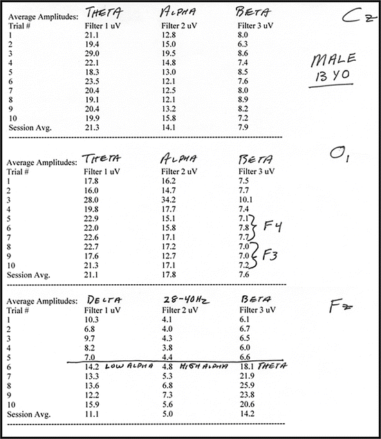
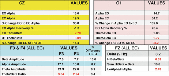

Fig. 2.5
Raw data from ClinicalQ recording of a 14-year-old male

Fig. 2.6
Summary statistics for the ClinicalQ shown in Fig. 2.5. Areas of diagnostic importance highlighted in red (Colour figure online)
We will be reviewing a great many data recordings of children with all manner of neurological issues that adversely affect their ability to pay attention and to learn. We will also be examining a great many records of children whose problems are emotional and behavioral in nature and not primarily the result of neurological problems. For the present purposes, the basic data recording will be reviewed to illustrate how profoundly accurate and sensible the ClinicalQ EEG method is, relative to the ubiquitous top-down methods. The actual data calculations are also presented so the reader can appreciate how straightforward and uncomplicated the procedure is for obtaining the ClinicalQ. The specific items that facilitated the precision of diagnosis will be identified but just the essentials that guided the evaluation of this child’s presenting complaints. These essential indicators are circled in red to set them apart from the other summary statistics.
As we proceed through the book looking at a number of different cases, the significance of all of these summary numbers will become apparent. For the present purposes, we want to focus only on those that have been highlighted, to demonstrate the remarkable efficiency of allowing the brain to tell us what the problems are, and where to go to fix them. The first indicators are at location Cz, directly on top of the head. The first number, 2.70, is the ratio of the amplitude of Theta (brainwaves from 3 to 7 cycles per second) divided by the amplitude of Beta (brainwaves from 16 to 25 cycles per second). The Theta/Beta ratio is extremely important in that it gives us an indication of the level of arousal of specific areas of the brain. We have databases for normative values for the normal functioning brain. The Theta/Beta ratio at that location for a child of about 14 years of age should be below about 2.20. Mitch’s ratio is 2.70. What this tells us is that this child has some difficulty associated with focus. When that area of the brain is hypoactive as indicated by elevated Theta/Beta ratio, there is too much slow activity and/or too little fast activity. This indicates that Mitch is facing a challenge in terms of focus, concentration, attention, and staying on target. If that ratio was considerably greater, up in the range of four or so, we would likely be probing to determine if Mitch is hyperactive. However, in the present case it is more likely that Mitch’s ADHD is of the inattentive variety.
What is most critical in this particular profile is the second number on that line, which is 3.09. This is the Theta/Beta ratio that was obtained when the child was under cognitive challenge. This was done during the time that he was asked to read aloud. Notice that the number increased from 2.70 to 3.09.
This is a particularly pernicious form of ADHD. When under cognitive challenge such as reading, the brain should be producing less slow frequency (i.e., lower amplitude or strength) associated with hypoactivity and/or greater fast activity associated with focus and attention. When it goes the opposite way (the ratio of the amplitude of slow frequency vs. high frequency gets larger), then this is a condition in which the harder the child tries, the worse the situation gets. We tend to find this condition mostly in males. The curious feature of this form of ADHD is that in some clients there are conditions in which the brain looks absolutely fine. The only time one sees the anomalous brainwave activity is when the child is being cognitively challenged. Thus, measuring brainwave activity when the child is simply sitting and not engaged does not reveal the condition that is causing the problems. Only when the child is asked to read aloud, or to count, do we see the elevated slow frequency amplitude.
The person who discovered this form of ADD is Professor George Fitzsimmons of the University of Alberta. The number of children who show the pattern just described (only see ADHD EEG profiles when being cognitively challenged) is not large. In most cases, one also sees neurological ADHD patterns even when the child is at rest. The important feature of this condition, however, is that cognitive challenge intensifies the condition. The usual result of this is that the harder the child tries, the worse the situation becomes. When trying to concentrate, the brain is showing greater amplitude of a brainwave that is associated with daydreaming and early stages of sleep. The tragic result is that children like this are highly at risk for simply giving up. They make determined efforts to keep up, and despite these efforts, they fall behind. These kids conclude that they are stupid or deficient in some way and simply give up. The giving up may have the form of rebellion, aggressive behavior, defiance and the like, or simply withdrawal. And our prisons are overloaded with the casualties of this condition.
Moving on to F4 and F3, we see the Theta/Beta ratios are 2.94 at F4, which is the right frontal cortex, and 3.04 at F3, which is the left frontal cortex. Whenever we see elevated slow frequency or elevated Theta/Beta ratio over the sensory motor cortex (i.e., location Cz), we typically see it as well in the frontal cortex. Elevated Theta/Beta ratio in the frontal cortex is associated with hypoactivity of these regions of the brain and reflected in some inefficiency in cognitive processing.
So the first thing I know about this child is that he has a pernicious form of ADHD. In general, I know that the child has likely made efforts to try to pay attention and do his homework. However, he finds that no matter how hard he tries, the problems simply seem to get worse.
There are several other “flags” in Mitch’s ClinicalQ that we will attend to shortly, but at this point I have enough information from the three circled areas (CZ, F3, and F4) to be able to discuss the situation with the child in front of me. So I say to, Mitch, “Mitch what the brain is telling me is that you have some problems staying focused in class. You find it difficult to pay attention, your mind tends to wander, and you have the same kind of problem when you try to do homework.”
I now have Mitch’s attention—he’s focused on me. “But there is another thing in this record,” I continue, “that’s really problematic.” “And it always causes students a lot of difficulty.” “What the brain is telling me is that the harder you try the worse the situation gets.” “No matter how hard you try, most of the time you find it extremely difficult to stay focused and on target both in class and when you are trying to do homework.” “This is a problem we find mostly in men and it really makes you want to just give up!”
As is common at this point in my feedback to the child, Mitch is having difficulty maintaining composure. As I have been told by many children after their treatment is completed, they found that I was the “one person on the planet who understood” (to quote one recent client) what the situation was. They did not have to spend any time telling me what the problem was—I was able to see it from what the brain was telling me.
At this point I turned to Mitch’s mother and asked if she would mind if I spoke with Mitch privately for a few moments. I often do this with teenage male clients for I find that it provides an opportunity for getting the child on board and committed to therapy. This is an opportunity to speak with the child without parents interrupting making comments and preventing me from developing good clinical attachment and report with the child.
In this particular case I noted several features of Mitch’s brain assessment that made me suspicious about marijuana use. These indicators were elevated slow frequency Alpha and elevated slow frequency in the back of the brain under eyes-closed conditions. Very often you find this with individuals who are cannabis users. I decided to take a chance once I had developed some rapport with Mitch. Mitch and I spoke about the use of neurotherapy with professional sports teams and with the Canadian Olympic athletes. I then said: “Mitch it is important that you be part of your treatment team. I can help you with the brain inefficiencies that I see here in this brain map but it’s important that you do what is necessary for these treatments to be really effective. And what I want you to do is stop smoking dope. Don’t say ‘yes’ or ‘no.’ If you’re not smoking dope so much the better but I’m getting some markers in your brain map that are often associated with cannabis use. If you are, stop because it makes people stupid. What cannabis does to people in your age range is it slows down a really important waveform in the brain and we certainly don’t want that to happen.”
As it turns out I was correct. Mitch was experimenting with marijuana. Mitch was so shaken by the accuracy of the brainwave assessment that I think he was shocked into stopping the marijuana use on the spot. We had a number of conversations and he shared with me later that he really felt like just quitting. He tearfully related that no matter how hard he tried, he simply could not function efficiently in school. He had great difficulty staying on target, doing his homework and not “looking stupid.” He said he just couldn’t wait until he could stop going to school. Neurotherapy saved this child’s life, a sentiment expressed on several occasions by his mother.
This is the form of ADHD that, in my judgment, is the one form that is most represented in the statistics associated with ADHD and criminality. These are the kids that quit; these are the kids that become truant; these are the kids that act up in school; these are the kids that become marginalized; these are the kids that get themselves into trouble; and these are the kids that are associated with the statistics about the number of incarcerated youth that have the symptoms of ADHD.
So the 14-year-old young lad who came into my office in a sullen, bored, and clearly frightened state was indeed fortunate because the diagnosis and treatment of this child at this age clearly saved his life. Looking at the risk factors, it only makes sense to neurologically evaluate the condition of these children as soon as they run into difficulty in school. Teachers and parents are very aware of these behaviors very early in the child’s life. The ease with which we can assess and diagnose the neurological anomalies is such that it is a tragedy that we are not doing so in our school systems.
Mitch is going on 20 at the time of this writing and is in the first few weeks of the third year of his undergraduate studies. Mitch had a total of 33 sessions between the ages of 14 and 16 and came back for a few more treatments when he felt that he was struggling at University.
The basic procedure at location Cz is to record ten 15-s epochs during which the client is engaged in specific activities. The client remains quietly observing the screen for two epochs following which there is one epoch of eyes closed. After epoch three the client’s eyes are again open. It is important to be precise with the instruction to open and close the eyes. One wants to see rapid increase in Alpha and then sharp decrease in Alpha when the eyes are again opened. It is also important to watch the raw signal (or spectral display) to determine that the Alpha response is “healthy.”
The remarkable clinical data ranges, such as ratios and summated bands, should be considered in the context of client variables such as age. For example, young children would be expected to have higher Theta/Beta ratios than adults. Hence, particular ratio levels are indicated as guidelines for the clinician to consider probing the client regarding a specific problem or characteristic. The basic clinical probes associated with the clinical norms are presented in this chapter. (The summary guide for administering the ClinicalQ and for the suggested clinical probes is found in Appendix A).
The raw data obtained from the ClinicalQ contains a wealth of information. The basic clinical probes provide the information for the intake session. As described by Susan Olding, it will allow surprisingly accurate identification of the problems for which the client is seeking treatment. However, there are many subtleties and nuances in the data set that will become apparent as one gains experience with “reading the Qs.” In the following sections, in addition to the research supporting the clinical probes, some of the statistically significant nuances will be presented. These nuances often guide the clinician’s formulation of a more textured conceptualization of the patient’s situation.
Unremarkable Clinical Ranges
Location Cz
At this location, three conditions are needed: Eyes open (EO), eyes closed (EC), and cognitive challenge (e.g., reading or counting backwards). In my clinic we also use this opportunity to determine the efficacy of some home treatment items such as harmonic sounds that influence brainwave activity. The OMNI Harmonic (Swingle 2010) that suppresses Theta amplitude, for example, is usually tested at location Cz.
Unremarkable clinical ranges for the measurements at Cz include: Theta/Beta ratio below 2.20 during EO and during cognitive challenge; Theta/SMR ratio below 3.00; the increase in Alpha band amplitude should be at least 30 % EC relative to EO; Alpha should block rapidly (i.e., drop to EO level quickly) so the post-EC epoch should be close to the pre-EC epoch Alpha amplitude if the Alpha blocking is efficient; TA (i.e., the sum of the amplitude of Theta, Alpha, and Beta bands) should be below 60.
Location O1
As most readers will surmise, the selection of location O1 reflects the legacy of Peniston’s work on Alpha/Theta training (Peniston and Kulkosky 1999). At this location, the ClinicalQ is assessing the quietude of the CNS, the Alpha response, Alpha blocking, and excessively high, slow frequency amplitudes.
Unremarkable ranges are a Theta/Beta ratio between 1.80 and 2.20 both EO and EC, Alpha amplitude increase of at least 50 % EC relative to EO, return of Alpha amplitude to EO level within one epoch (i.e., the post-EC epoch is within 25 % of the pre-EC epoch amplitude), and TA below 60.
Locations F3 and F4
The ClinicalQ provides a wealth of information about behaviors from the assessment of the frontal cortex. Basically, the two locations should be balanced and the ratios among Theta, Alpha, and Beta to be symmetrical. The assessment of all frontal locations is done EC to reduce eye movement artifact problems. In my experience, a “rule-of-thumb” indicator of imbalance in the amplitudes in the frontal lobes is when the disparity is greater than about 15 %. However, as will be discussed below, we do find statistically significant differences in self-rated conditions when the disparity is 10 % or greater. These lower disparities may be conceptualized as shadow symptoms, but treatment of these imbalances is identical to those in the remarkable ranges.
In the frontal lobes, we want to see the amplitude of any brainwave band to be about the same at both F3 and F4. The unremarkable range would be that the difference between F3 and F4 in any band is less than 10 %. The Theta/Beta ratio is below 2.00 and the Theta/Alpha ratio is between 1.25 and 1.75. The TA is below 60.
Location Fz
At this location, the ClinicalQ assesses the activity of the anterior cingulate gyrus, the Delta amplitude, and the ratio of Lo-Alpha to Hi-Alpha.
Normative ranges at location Fz are Delta below 9.0, the ratio of 28–40 Hz amplitude to Beta amplitude between 0.45 and 0.55, the sum of the latter two bands below 15, and the ratio of Lo-Alpha to Hi-Alpha below 1.50.
Remarkable Ranges
Sample Population
The ranges suggested as normative are based on more than three decades of clinical observation. There may be some slight differences in the ranges depending on the type of clinical EEG systems one is using. The ranges and threshold values, then, are offered as starting points to aid clinicians in identifying the EEG signatures that are associated with clinical symptoms.
On the first visit, every client completes a series of forms including various consent forms for treatment, authorization forms, basic biographical information forms, and two forms that ask about symptoms and behaviors. These forms are shown in the Appendices at the back of this book. The data for the statistical analyses presented throughout this manual are drawn from these intake questionnaires.
The statistical data presented below are offered to validate the robust features of the ClinicalQ. The sample consists of clients seen in my Vancouver clinic over the last several years. Only clients with complete data sets were included in the data sample. Further, only clients who did not designate a head injury or a condition that necessitated an initial full QEEG were included. The sample therefore excludes clients with epilepsy, traumatic brain injury, Parkinson’s, stroke, Alzheimer’s, and the like. The adult sample consists of 732 clients. The child sample consists of 547 clients.
Specialty populations include seniors, over 80 years old (N = 30); left handed (N = 110); independently diagnosed (by psychiatrist) bipolar (N = 37); Sleep disordered with four-night EEG monitoring (N = 52). All samples are combined gender—no clinical benefit has been found for independent gender norms. Unless otherwise noted, all probabilities are based on 2-tail statistical distributions.
Clinical Implications of Remarkable Ranges
The above normative ranges were established from data collected on clinical populations. The normative values will vary as a function of many variables such as time of day, medication/drug history, physiological state of the client, and client age, to name but a few.
The reader may note that these normative values are far more stringent than those of the normative databases. Further, studies comparing clinical populations versus nonclinical populations (e.g., ADHD vs. nonclinical comparison group) find normative discriminative ranges that are considerably higher than some of the normative ranges of the ClinicalQ. The reason for this is obvious. Clients come to a neurotherapist complaining, for example, of problems with attention and focus. Relative to their own EEG, one may find that the only area showing any elevation is in the excess of Theta amplitude, reflected in the Theta/Beta ratio, over the sensory motor cortex (location Cz). This elevation may seem minimal with a ratio in the low 2 plus range. In any controlled study, comparing this person with a group of nonclinical cohorts, a ratio of that magnitude would probably not be statistically different from the control group mean. However, that does not mean that this is not the brain condition associated with this client’s complaint of problems with attention. One client with a Theta/Beta ratio of 2.50 at location Cz may not complain of any problems with focus and hence they do not appear in the neurotherapist’s office. Another person with exactly the same ratio, on the other hand, may seek treatment for attention problems and find that the condition improves when the ratio is reduced. In short, the remarkable ranges are clinical heuristic ranges. Whether or not they survive discriminative function statistical comparisons with nonclinical groups is largely irrelevant. One should always keep in mind that the client is sitting in the neurotherapist’s office because he or she has a troublesome symptom. The beneficial feature of the ClinicalQ is that one can isolate areas of brain activity that may be associated with the symptom even if the activity is not statistically different from a nonclinical group normative range.
Symptoms Associated with Remarkable Ranges at Location Cz
Two and one half minutes of recording at location Cz provides the neurotherapist with a wealth of valuable information. First, one looks at the overall Theta/Beta ratio. Ideally, we want to see that ratio below 2 or so. If the ratio is above 2.20, probe the client to determine if focus and attention are a problem. Even if the overall, or mean, ratio is below 2.20 but one finds that under cognitive challenge (e.g., reading, counting) the ratio goes above 2.20, also probe for attention problems.
From our clinical sample, of the 39 children who were rated by a parent as “hyperactive” (4 or 5 on a five-point scale), 71.8 % had Theta/Beta ratios above 2.20 and 28.2 % had ratios below 2.20 (z = 4.27, p < 0.001, N = 39). Sixty-seven children from our sample were rated by a parent as 4 or 5 on the item “attention problems in school.” From this sample, 70.2 % had a Theta/Beta ratio above 2.20 and 29.8 % had ratios below 2.20 (z = 5.11, p < 0.001, N = 67). The finding of elevated Theta/Beta ratios in clients diagnosed with ADHD is quite robust. Thompson and Thompson (2006), Chabot et al. (2001), and Barry et al. (2006) likewise report that a large proportion (around 80 %) of clients diagnosed with ADHD show this elevated Theta/Beta ratio pattern. Even minor elevation in the Theta/Beta ratio (above 2.30) results in a highly reliable increase in the average rating on the “problems with concentration” question (difference = 6.1 %, N = 631, t = 4.45, p < 0.001) and “…often do not remember what I have just read” question (difference = 3.8 %, N = 635, t = 2.35, p < 0.02).
If the Theta/Beta ratio jumps substantially (25 % or greater) under a cognitive challenge, such as reading, then probe to determine if the client frequently experiences reading a paragraph and then discovering that they cannot remember what they had just read. In my experience, one usually gets an affirmative reply to that query.
From our adult database, 59 clients’ intake data showed at least a 15 % jump in the Theta/Beta ratio under reading condition. Of that sample, 6.8 % rated themselves as not experiencing problems with remembering what they have just read whereas 45.8 % of this sample of adults rated themselves high on this same questionnaire item (z = 5.34, p < 0.001, N = 59).
It appears that this form of ADHD, when the Theta/Beta increases under cognitive challenge, is a particularly pernicious form. Experience with many clients gives the clinical impression of greater struggle and discouragement with academic work. One gets the impression of children, in particular, wanting to just give up and quit because “the harder they try the worse the condition gets,” a statement that always seems to resonate with these children. It is important to also note that, at rest, this condition is frequently missed because the ratios remain within normal range. It is only when the child is cognitively challenged with reading or a mental math task that the condition manifests.
Related findings seem relevant here. Parent rating of “easily frightened” is related to the Theta/Beta ratio under challenge. Children rated, by a parent, as “easily frightened” had an, under challenge, Theta/Beta ratio that was, on average, 10 % greater than the at rest ratio (t = 2.33, p = 0.02, df = 143). This relationship between fear and the Theta/Beta ratio was not found for the at rest condition. Also, children rated as “Stubborn” had under challenge Theta/Beta ratios that were, on average, 15 % greater than those rated low on this trait (t = 5.60, p < 0.001, df = 162). This relationship also was not significant under at rest conditions.
If the Theta/Beta ratio remains relatively constant between rest and cognitive challenge but one observes that the Beta has increased substantially (greater than 25 %) between these two conditions, then probe the client to determine if they find reading fatiguing. Often we find that the Theta amplitude increases substantially under cognitive challenge, but this increase is not reflected in an increased Theta/Beta ratio because of a simultaneous increase in Beta amplitude. It appears as though the client is compensating for the Theta amplitude increase by increasing mental effort. This condition can also result from eye problems in which eyestrain is causing an EMG artifact resulting in increased amplitudes for both Beta and Theta.
If the Theta/Beta ratio is below 2.20 on average but well above that value when reading, this could be either ADD or have problems with comprehension/retention of information. Often, of course, it is both of these conditions. However, one does encounter clients for whom attention in class does not seem to be a major problem, but the children have considerable difficulties in comprehension and/or retention of written material.
Another condition that also is often associated with poor retention of written material is when the Alpha response is blunted or absent. The increase in Alpha amplitude should be at least 30 % between eyes open and eyes closed. When the Alpha response is deficient, clients often complain of poor retention of information and/or poor short-term memory. On the simple question of “I am forgetful” clients with an Alpha response below 15 % at Cz had an average response 8 % higher (more forgetful) as compared with clients with Alpha responses above 35 % (t = 2.36, df = 640, p < 0.02). Those with an Alpha response greater than 35 % were more than twice as likely to rate themselves as having no problem at all with forgetfulness (1 on the 5-point scale) as compared with those with an Alpha response below 15 % (z = 2.59, p < 0.01, N = 643). Conversely, those rating themselves as forgetful (4 or 5 on the 5-point scale) versus those who rated themselves as not forgetful (1) had an Alpha response that was, on average, 28.2 % lower in amplitude (t = 2.06, p = 0.04, df = 380).
Slow return of the Alpha response (Alpha amplitude return to eyes-open level following eyes-closed condition) was related to children’s “sleep disturbances” as rated by a parent. The Alpha amplitude level of the sleep disturbed rated child was 117.6 % greater than the child without a sleep problem (t = 2.12, p < 0.04, df = 166). This could indicate that the sleep disturbed child has a problem shutting off visual images or perhaps more difficulty in state changes. Although no data are available, clinical impression also suggests that some individuals with a history of emotional trauma also show slow Alpha returns as well.
When the Alpha response is blunted or negative (i.e., the Alpha amplitude is lower under eyes-closed condition than under eyes-open condition) then one should consider the possibility of traumatic stress (Swingle 2001, 2013). If the Alpha response is negative at both locations Cz and O1, there is a strong likelihood that the client has experienced or is experiencing emotionally traumatic conditions.
I became aware of this trauma signature when working with clients with Posttraumatic Stress Disorder (PTSD) at McLean Hospital. These traumatized clients included Vietnam veterans and police officers, among others. It became obvious during the ClinicalQ that these clients had no Alpha response at Cz and/or O1. There were a few exceptions, as will be discussed below, but the vast majority of the clients that I treated showed this deficit. Metaphorically, one can conceptualize this as the brain protecting itself against the major stressor of PTSD, the flashbacks. Alpha being a visualization response, it is intuitively consistent that this dampened Alpha response would be reinforced by fear or avoidance.
If the Alpha response is negative at Cz but present at O1, then one occasionally finds that the client experiences memory and retention difficulties but does not admit to traumatic stress. In addition, when the blunting is only found at Cz, the emotionally stressing situation is more likely to be current or recent. Blunting that only appears at O1, on the other hand, is more likely to be associated with historical traumatic events. Again, these generalizations offer hypotheses for the clinician to probe with the client. However, as will be discussed latter in this book, these subtleties can offer remarkable insight into understanding the conditions that are affecting the client.
For example, in the Clinic, we often encounter children who have been referred for treatment of AD(H)D who show no EEG anomalies associated with attention or focus problems, but show the trauma signature. Proceeding cautiously, we probe for past or present emotionally traumatic stress. We have encountered a number of cases in which the child’s attention and focus problems were stress related and not a function of any AD(H)D brain pattern. Several examples of recent cases include bullying, hospital experiences, serious parental illness, sexual abuse, and family violence. Later in this book many such cases will be reviewed.
As one would expect, individuals with these trauma markers frequently report depressed and/or anxious mood states. From our database, clients who rated themselves at level 4 or 5 (on a five-point scale) on “I feel depressed” had Alpha responses almost 50 % lower in amplitude on average (48.8 μV vs. 72.4 μV; n = 346, t = 3.09, p < 0.004) as compared to those rating themselves at a 1 (“Not true of me”). Those rating themselves at 4 or 5 on “I am very anxious” had an Alpha response that on average was 27.3 % lower in amplitude (51.6 μV vs. 65.7 μV; n = 437, t = 2.14, p < 0.04) than those rating themselves at a 1.
There are many issues that must be addressed with respect to how one should proceed when the trauma signature is encountered. First, is it really trauma? We have encountered situations with both adults and children in which the clients were not aware of, or did not admit to, any trauma. In my experience this is relatively uncommon, although we have encountered clients with severe learning disorders, and markedly blunted Alpha, who are adamant that trauma was not part of their history.
Second, and most importantly, is the neurotherapist competent to deal with trauma and, if not, is the neurotherapist properly networked to refer the client to a competent person? Neurotherapy is not a stand-alone discipline and nowhere is this more apparent than in the case of trauma. Neurotherapists without adequate psychological training should not handle cases of this nature. At best, the client is not going to improve much, because a major cause of the client’s difficulties is left unaddressed. At worst, the client is going to abreact with a neurotherapist not equipped to deal with problems of this severity.
Drawing examples from our adult database, when the Alpha response is below 10 % at both Cz and O1, over 80 % admit to having a history of emotional trauma (z = 4.40, p < 0.001, N = 54). The situation is not as clear when dealing with individuals with a strong Alpha response, such as a person with strong visualization skills (the artist signature).
Strong Alpha response at both locations Cz and O1 has been shown to be related to clients rating themselves as “artistic.” Clients with Alpha responses above 80 % rated themselves as high on the “I am very artistic” questions, 47.3 % more often than clients with Alpha response below 25 % (z = 2.86, p < 0.005, N = 591). Clients rating themselves high on the artistic question (4 or 5) had an Alpha response that was 26.1 % stronger, on average, as compared with clients who rate themselves at a 1 level (t = 2.08, p < 0.04, N = 415). This “artists’ signature” is also found at location O1 as will be discussed later. It is interesting to note that those clients rating themselves as high on the artistic question tend to have higher Theta/Beta ratios at location Cz under eyes open (M = 8.9 % higher, t = 2.66, p < 0.01) and reading conditions (M = 8.2 %, t = 2.33, p < 0.02).
When clients have strong resident Alpha (e.g., artists), the blunting effect of trauma can be obscured in the summary statistics. Skilled neurotherapists can see trauma on a strong Alpha response because of the unique patterning. The “traumatized artist signature” on the raw signal or EEG single hertz spectral display has the look of the Alpha amplitude being “pushed down.” It is also frequently found that current exposure to severe emotional stressors with clients who have the artist’s strong Alpha response will show blunting at Cz and much less so at O1. This was the pattern observed in the previously discussed case of the child who was being bullied at school. His Alpha response at location Cz was about 19 % whereas at O1 it was nearly 100 %.
A related finding also suggests that the blunted Alpha response is related to exposure to emotional trauma and further that this exposure may have effects on the child’s sense of security. Children whose parents rated them as “easily frightened” had an average Alpha response of 27.6 % (below the clinical threshold for trauma marker) as compared with an average response of 49.5 % for children rated as not easily frightened (t = 3.39, p < 0.001, df = 152). This is directly relevant to the issue of causes other than ADHD that effect a child’s academic performance. They may just be afraid, hypervigilant, or insecure which in turn affects their ability to focus and be attentive in school. The case of the child with a ClinicalQ profile suggesting that he may have been the victim of bullying, discussed in this book, is a good example of a situation in which a frightened child was assumed to have ADHD when in fact the fear was a primary cause of his problems with focus.
Two cases of clients who experienced serious trauma during the time of their treatment with us show how trauma suppresses the Alpha response. Both of these clients had strong Alpha responses, and both rated themselves as having good artistic skills on our intake questionnaires. In one case, a young child found a dead body while playing in a wooded area, and the second case was a young woman whose best friend committed suicide just one day before we remapped her Alpha response. In the first situation, the Alpha response was reduced by 69.4 % after the trauma. The treatment for this condition, in addition to counseling, is to “release” the Alpha response with neurotherapy and/or emotional release therapies including EMDR, somatoemotional release, hypnosis, and the like (Swingle 2006, April). In this case, neurotherapy was all that was required, in addition to the counseling. After three neurotherapy sessions of Alpha increase at Cz and O1, the Alpha response was restored to within 12 % of the pre-trauma level. In the second case mentioned above, this 20-year-old young woman learned of the suicide of her best friend just one day before the brain assessment. The Alpha suppression was 21.9 % in one location and 46.5 % in the second location.
Research from the Swingle Clinic (Swingle 2013) indicated that the Alpha response could be blunted with brief exposures to strong emotionally negative photographs. Clients viewed either no pictures, a positive picture (horse and farmer in a field), or a negative photo (dead body being dragged with an ice hook on the skull in Nazi concentration camp) for 10 s. The Alpha response taken before viewing the pictures (or no picture control) was compared with a second Alpha response after the experimental condition. The negative photo resulted in an average Alpha response blunting of −62.3 %. The positive photo and no photo viewed conditions both resulted in the second Alpha response being greater than the first (no photo = +33.3 %; positive photo = +108.9 %; (all comparisons with negative photo, p < 0.0005).
Stay updated, free articles. Join our Telegram channel

Full access? Get Clinical Tree


