CHAPTER 19
The Limbic System
The limbic system subserves basic survival functions that include feeding behavior, “fight-or-flight” responses, aggression, and the expressions of emotion and of the autonomic, behavioral, and endocrine aspects of the sexual response. It includes phylogenetically ancient portions of the cerebral cortex, related subcortical structures, and fiber pathways that connect with the diencephalon and brain stem (Tables 19–1 and 19–2).
Table 19–1 Components of the Limbic System and Neocortex.
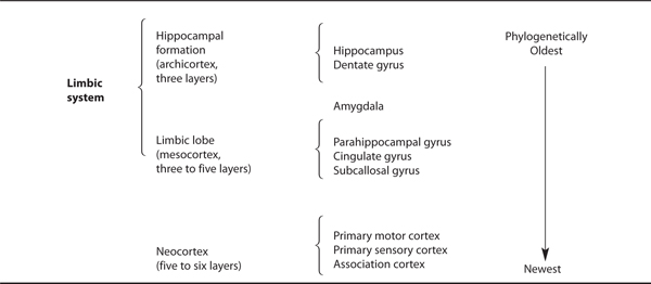
Table 19–2 Major Limbic System Connections.
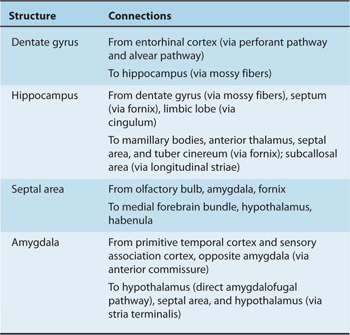
The limbic system receives input from many parts of the cortex and contains multimodal association areas where various aspects of sensory experience come together to form a single experience. The hippocampus, within the limbic system, plays crucial roles in spatial problem solving and in memory.
THE LIMBIC LOBE AND LIMBIC SYSTEM
The limbic lobe was so named because this cortical complex forms a limbus (border) between the diencephalon and the more lateral neocortex of the telencephalic hemispheres (Fig 19–1). This limbic lobe consists of a ring of cortex outside the corpus callosum, largely made up of the subcallosal and cingulate gyri as well as the parahippocampal gyrus (Fig 19–2).
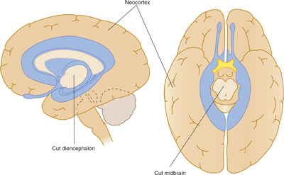
FIGURE 19–1 Schematic illustration of the location of the limbic system between the diencephalon and the neocortical hemispheres.
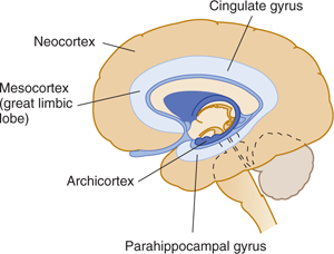
FIGURE 19–2 Schematic illustration of the concentric main components of the limbic sytem.
More recent authorities revised the concept of the limbic lobe and refer to the limbic system, which includes the functionally interrelated limbic lobe (parahippocampal, cingulate, and subcallosal gyri), the amygdala, and the hippocampal formation and associated structures (see Table 19–1). The hippocampal formation (a more primitive cortical complex) is situated even closer to the diencephalon and is folded and rolled inward so that it is submerged below the parahippocampal gyrus. The hippocampal formation consists of the hippocampus (Amnion’s horn); the dentate gyrus; the supracallosal gyrus (also termed the indusium griseum), which is the gray matter on top of the corpus callosum; the fornix; and a primitive precommissural area known as the septal area (Fig 19–3).
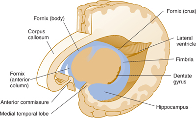
FIGURE 19–3 Schematic illustration (left oblique view) of the position of the hippocampal formation within the left hemisphere.
Histology
The cortical mantle of the brain can be thought of as consisting of three concentric cortical regions (hippocampal formation, limbic lobe, and neocortex) with different cytoarchitectonic features (Fig 19–4). The innermost of these three regions, the hippocampal formation, is the most primitive, and the outermost, the neocortex the most advanced. The most primitive cortex, which constitutes the hippocampus, also termed the archicortex, has three layers. The cortex of the transitional limbic lobe—the mesocortex, or juxtallocortex—has as many as five layers. The remaining cortex, known as the neocortex, or isocortex, is phylogenetically newest and has five or six layers. It includes the primary motor and sensory cortex as well as the association cortex and covers most of the cerebral hemispheres (see Chapter 10).
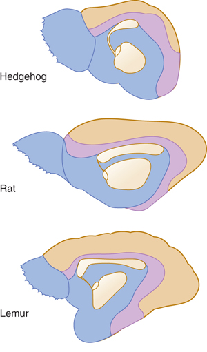
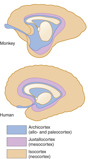
FIGURE 19–4 Diagrams of the medial aspect of the right hemisphere in five species. Note the relative increase in size of the human neocortex (isocortex).
The concentric architecture is more obvious in lower species. It is also present in higher species (including humans) and underscores the tiered arrangement of a phylogenetically advanced neocortex, which rests on a more primitive and deeply buried limbic lobe and hippocampal formation. Because of their purported role in olfaction, the hippocampal formation and limbic lobe were also termed the rhinencephalon (“smell-brain”) by classical neuroanatomists. More recent work has shown that limbic structures are related to the sense of smell but are also directly involved in primitive, affective, visceral, and autonomic functions. Such names as the visceral brain, emotional brain, and limbic brain were once used but were discontinued in favor of the more neutral limbic system.
OLFACTORY SYSTEM
Olfaction (the sense of smell) is one of the oldest senses from a phylogenetic point of view. The olfactory system constitutes an important input to the limbic system, which is also phylogenetically old.
Olfactory Receptors
The olfactory receptors are specialized neurons located in the olfactory mucous membrane, a portion of the nasal mucosa. The olfactory mucous membrane is blanketed by a thin layer of mucus, produced by Bowman’s glands. The olfactory receptors are highly sensitive and respond with depolarizations when confronted with odor-producing molecules that dissolve in the mucous layer. The olfactory receptors contain, in their membranes, specialized odorant receptors that are coupled to G-protein molecules, which link these receptors to adenylate cyclase. There are nearly 1000 odorant receptor genes; each olfactory receptor expresses only one or a few (and thus responds to only one or a few odoriferous molecules). When a specific odoriferous molecule binds to the appropriate olfactory receptor, it activates the G-protein molecule, which, via adenylate cyclase, generates cyclic adenosine monophosphate (AMP); this, in turn, leads to opening of Na+ channels, generating a depolarization in the olfactory receptor.
The axons of the olfactory receptors travel within 10 to 15 olfactory nerves to convey the sensation of smell from the upper nasal mucosa through the cribriform plate to the olfactory bulb (Figs 19–5 and 19–6). The olfactory bulb and olfactory tract (peduncle) lie in the olfactory sulcus on the orbital surface of the frontal lobe. As the tract passes posteriorly, it divides into lateral and medial olfactory striae (Fig 19–7). Within the olfactory bulb, the olfactory receptor axons terminate in specialized synaptic arrangements (termed glomeruli) on the dendrites of mitral cells (see Fig 19–6). Olfactory neurons expressing a specific odorant receptor (and thus responsive to a specific odorant stimulus) project precisely to a small number of glomeruli within the olfactory bulb. There thus appears to be a spatial map within the olfactory bulb that identifies the receptors that have been stimulated.
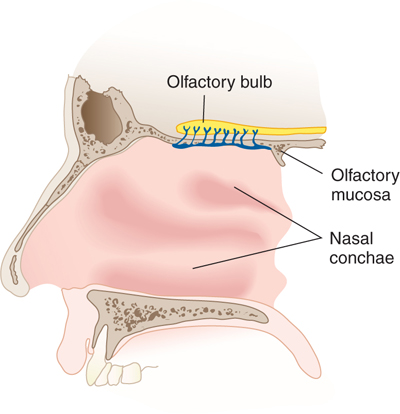
FIGURE 19–5 The olfactory nerves (lateral view).
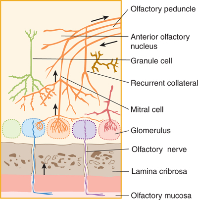
FIGURE 19–6 Neural elements in the olfactory bulb.
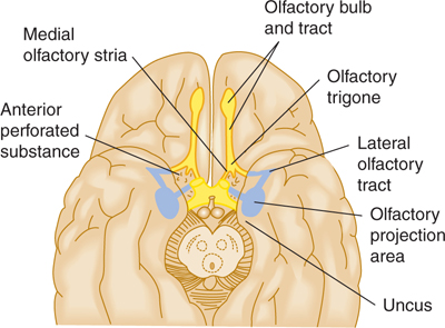
FIGURE 19–7 Olfactory connections projected on the basal aspect of the brain (intermediate olfactory tract not labeled).
The mitral cells of the olfactory bulb send their axons posteriorly via the olfactory tracts (also termed the medial and lateral olfactory stria) to the olfactory projection area in the cortex. The lateral olfactory stria is the projection bundle of fibers that passes laterally along the floor of the lateral fissure and enters the olfactory projection area near the uncus in the temporal lobe (see Fig 19–7).
The olfactory projection area is the part of the cortex that receives olfactory information. The olfactory projection area includes the pyriform and entorhinal cortex and parts of the amygdala. The pyriform cortex projects, in turn, via the thalamus to the frontal lobe, where conscious discrimination of odors presumably occurs.
The small medial olfactory stria passes medially and up toward the subcallosal gyrus near the inferior part of the corpus callosum. It carries the axons of some mitral cells to the anterior olfactory nucleus, which sends its axons back to the olfactory bulbs on both sides, presumably as part of a feedback circuit that modulates the sensitivity of olfactory sensation. Other olfactory fibers reach the anterior perforated substance, a thin layer of gray matter with many openings that permit the small lenticulostriate arteries to enter the brain; it extends from the olfactory striae to the optic tract. These fibers and the medial stria serve olfactory reflex reactions.
HIPPOCAMPAL FORMATION
The hippocampal formation is a primitive cortical structure that has been “folded in” and “rolled up” so that it is submerged deep into the parahippocampal gyrus. It consists of the dentate gyrus, the hippocampus, and neighboring subiculum.
The dentate gyrus is one of the few regions of the mammalian brain where neurogenesis (the production of new neurons) continues through adulthood.
CLINICAL CORRELATIONS
Related posts:

Stay updated, free articles. Join our Telegram channel

Full access? Get Clinical Tree


