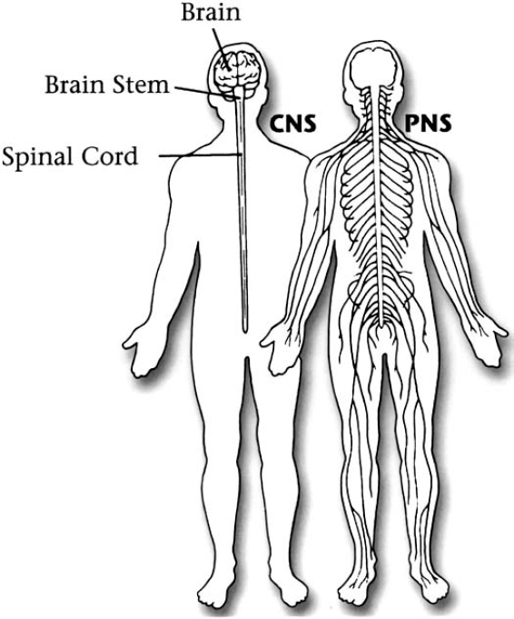
2 The diagnosis of neurofibromatosis is based on clinical criteria established mainly through a detailed medical history and physical examination. Although other varieties of neurofibromatosis may exist, genes have been found, and diagnostic criteria established through consensus committees, for only two: neurofibromatosis 1 (NF1) and neurofibromatosis 2 (NF2). Criteria have been proposed for a third type of neurofibromatosis, schwannomatosis, as the search for its cause continues. The current criteria to diagnose NF1 and NF2 were issued in 1997 by the National Neurofibromatosis Foundation Clinical Care Advisory Board.1 The board not only reviewed and updated diagnostic criteria and management guidelines issued originally in 1987, and updated in 1990, by the National Institutes of Health,2,3 but also conducted its own independent review of relevant research studies. In most cases, the criteria detailed in Chapters 5, 10, and 12 can be used to establish or rule out a diagnosis of neurofibromatosis. Because many features of these disorders are age dependent, however, diagnosis may take several years, especially if there is no family medical history of neurofibromatosis. Although this may frustrate patients and their families, at this point there are few alternatives to a strategy of watchful waiting. No laboratory test establishes the diagnosis, although sometimes tissue biopsies or radiological films, such as magnetic resonance imaging (MRI) scans and x-rays, provide additional information to help confirm one. Even though the genes for both NF1 and NF2 have been identified and clinical genetic testing is available, it is not appropriate for everyone and in most cases does not influence management of the disorders (see Chapter 4). Nor is there a reliable blood test: the NF1 and NF2 proteins both function inside cells and therefore cannot be measured in the same way as blood sugar, cholesterol, and other biological products that circulate in the bloodstream. The responsible mechanism for schwannomatosis has not yet been identified and no genetic test exists. Although it is human nature to want to know something definitive, it is important to remember that in most cases early diagnosis of neurofibromatosis does not have an effect on management of these disorders. The most productive strategy, for physicians and patients alike, is to adopt an attitude of informed vigilance. (This chapter and the ones that follow provide information to facilitate doing just that.) The neurofibromatoses are primarily evident in cells and connective tissue in the nervous system, but they are otherwise distinct. Given their significantly different clinical manifestations, it is somewhat surprising to think that NF1 and NF2 were once thought of as a single entity. Schwannomatosis is somewhat harder to distinguish from NF2 because the two disorders share a hallmark tumor type. Table 2–1 presents a quick summary of the differences between NF1, NF2, and schwannomatosis. Tumors in neurofibromatosis grow from cells and tissues that play essential roles in the functioning of the nervous system (Fig. 2–1). NF1 mainly affects nerve sheath cells in the peripheral nervous system. Its hallmark features include multiple café-au-lait spots and neurofibromas. The signature manifestation in NF2, which mostly is revealed in the central nervous system, is bilateral schwannomas that grow on the vestibular nerve, which connects the inner ear to the brain. Schwannomatosis was once considered a subtype of NF2 because it also causes multiple schwannomas to grow. In schwannomatosis, however, the tumors grow anywhere except on the vestibular nerve. Although it is possible that other types of neurofibromatosis exist, this is difficult to determine with certainty because these disorders vary widely in symptoms and severity. The cells that compose the nervous system are unique in that they form large networks and interdependent circuits that snake beneath the skin to every point in the body. Neurons, individual nerve cells in the brain and spinal cord, rely on a vast network of connections with other nerve cells to help people sense, move, and think. Nerve cells are connected by axons, which transmit electrical impulses much like wires in a house.
The Many Faces of Neurofibromatosis
♦ Common Features of Neurofibromatosis
| Typical Characteristic | NF1 | NF2 | Schwannomatosis |
| Gene location | Chromosome 17 | Chromosome 22 | Unknown but likely chromosome 22 |
| Onset of manifestations | Early childhood | Early adulthood | Early adulthood |
| First manifestations | Café-au-lait spots | Hearing and/or balance problems | Pain |
| Eye manifestations | Lisch nodules | Posterior subcapsular cataracts | None |
| Common developmental aspects | Large head Short stature Learning disabilities | None | None |
| Most common tumors | Neurofibromas Optic gliomas | Schwannomas (especially vestibular) Meningiomas | Schwannomas (any except vestibular) |
In neurofibromatosis, tumors primarily develop in support cells that surround nerve cells and help them to function. The cells most often involved are those that help to form protective insulation around axons known as the myelin sheath. In the brain and spinal cord, oligodendrocytes help to form myelin; in the peripheral nervous system Schwann cells perform this role. Connective tissue in the nervous system contains fibroblasts and meningeal cells. Astrocytes clean up excess transmitters and ions (which help neurons to communicate) and provide glucose to fuel neuronal activity. All of these cells have been found in various types of tumors caused by neurofibromatosis.
♦ Distinguishing Neurofibromatosis 1 from Neurofibromatosis 2
Although the consensus criteria and the discovery of two separate genes make it clear that NF1 and NF2 are two distinct disorders, the two are still sometimes confused at diagnosis. Most often, this occurs when a person with NF2 also develops some of the features that characterize NF1, notably skin tumors and café-au-lait spots. As Table 2–2 makes clear, however, the manifestations of NF1 and NF2 are sufficiently different that the two can be distinguished by careful physical examination and testing.
Related posts:
 Cognitive Function and Learning Disabilities in Neurofibromatosis 1
Psychosocial Impact of Neurofibromatosis
Genetics and Genetic Counseling in Neurofibromatosis 1 and Neurofibromatosis 2
The Pathogenesis of Neurofibromatosis 1 and Neurofibromatosis 2
Other Features of Neurofibromatosis 1
Neurofibromas and Malignant Peripheral Nerve Sheath Tumors (MPNST) in Neurofibromatosis 1
Cognitive Function and Learning Disabilities in Neurofibromatosis 1
Psychosocial Impact of Neurofibromatosis
Genetics and Genetic Counseling in Neurofibromatosis 1 and Neurofibromatosis 2
The Pathogenesis of Neurofibromatosis 1 and Neurofibromatosis 2
Other Features of Neurofibromatosis 1
Neurofibromas and Malignant Peripheral Nerve Sheath Tumors (MPNST) in Neurofibromatosis 1
Stay updated, free articles. Join our Telegram channel

Full access? Get Clinical Tree


