The Middle Cerebral Artery
Key Points
Variants of the middle cerebral artery, such as accessory branches or duplications, represent a substantial risk for misadventure during endovascular embolization or blind navigation during treatment of ischemic stroke. Probe gently.
The AP angiographic view can be very misleading as to the state of perfusion of the middle cerebral artery. Always review the DSA runs well into the parenchymal and venous phases, particularly on the lateral view, to search for late filling or slowly draining vessels, an indicator of distal embolic complications.
The vascular territory of the middle cerebral artery includes some of the most eloquent cortical areas for motor and sensory functions. It encompasses the receptive and expressive components of language, abstract thought, and other faculties of higher cognitive functioning. In addition, perforating branches of the proximal middle cerebral artery supply the basal ganglia and important descending and corticospinal tracts.
Segmental Anatomy of the Middle Cerebral Artery
The M1 (horizontal/sphenoidal) segment of the middle cerebral artery extends from the bifurcation of the internal carotid artery to the limen insulae, where the middle cerebral artery makes a turn or genu superiorly into the insula (Fig. 12-1). The M2 (insular) segment extends from the genu to the circular sulcus of the insula, and the M3 (opercular) extends from the circular sulcus to the opercular turn of the middle cerebral artery branches. The M4 (cortical) segments are those visible on the lateral convexity of the hemisphere (1,2).
Branching Patterns of the Middle Cerebral Artery
The middle cerebral artery can divide in up to four described patterns: a single trunk with no main division, a bifurcation, a trifurcation, or a quadrifurcation. Of these, the most common is a bifurcation pattern seen in 64% to 90% of hemispheres (2,3,4). Most neurology literature presupposes this pattern (5), and the terminology of superior and inferior divisions of the middle cerebral artery is in ubiquitous clinical use. A trifurcation pattern may be seen in 12% to 29% of hemispheres, with other patterns being less common.
Variability within the bifurcation pattern can be evident at microanatomical examination and in the clinical syndromes associated with a division occlusion. It is slightly more common (32%) for the inferior division to be dominant, covering a more extensive cortical area than the superior division. The superior division is dominant in 28% of hemispheres (Fig. 12-2). Balanced division is seen in approximately 18% of hemispheres (2), in which cases, the superior division spans the orbitofrontal to the posterior parietal areas.
Perforating Branches
The anterior perforated substance is the flat surface at the base of the brain bound medially by the interhemispheric fissure, laterally by the limen insulae, anteriorly by the division of the olfactory tract into the medial and lateral olfactory striae, and posteriorly by the optic tract and temporal lobe. It is divided along the sagittal plane into medial and lateral components by a line drawn posteriorly from the axis of the olfactory tract (6). This line divides the medial from the lateral anterior perforator or lenticulostriate vessels, which penetrate the perforated substance.
There are between 1 and 21 lenticulostriate branches per hemisphere, with an average of 10 (6,7,8). Approximately 80% arise usually from the posterosuperior aspect of the artery proximal to the middle cerebral artery division. The remainder arises from the proximal branches of the middle cerebral artery, particularly from the superior division (Fig. 12-3).
The lateral lenticulostriate arteries have a slightly larger diameter than the medial group. The lateral group in particular describes a recurrent curve before entering the anterior perforated substance. The lateral lenticulostriate arteries arise from the M1 or M2 segments of the middle cerebral artery. On AP projection, they have an S (on the right side) or reverse S (on the left side) shape. From their origin, they follow a sharp posterior and medial turn in the cisternal segment to assume a more lateral curve as they enter the anterior perforated substance. Here, they initially course around the lateral aspect of the putamen and fan out to give an important supply to the lateral aspect of the anterior commissure, internal capsule, dorsal aspect of the head of caudate nucleus, putamen, lateral globus pallidus, and substantia innominata. Blood supply to more lateral structures including the claustrum and external capsule derives from insular branches.
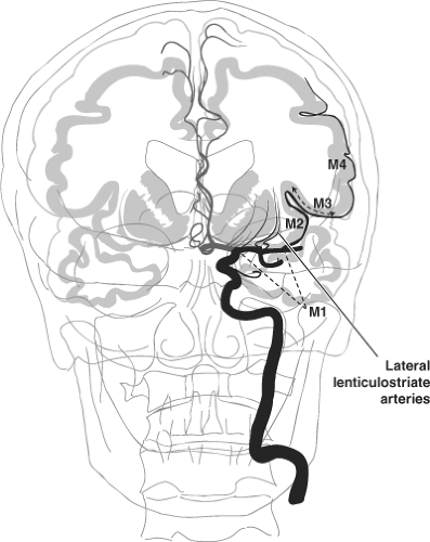 Figure 12-1. Segmental anatomy of the middle cerebral artery. The segmental anatomy of the middle cerebral artery is illustrated. Although the M1 segment was originally defined as extending from the carotid bifurcation to the limen insulae, in clinical parlance, the term M1 is most frequently applied to the main stem of the middle cerebral artery extending to the principal division. |
More than 70% of all aneurysms of the middle cerebral artery occur at its division, while aneurysms of the M1 trunk of the middle cerebral artery represent 10% of MCA aneurysms. They tend to arise either from the superior wall of the artery, in which case they are associated with the lenticulostriate arteries, or from the inferior surface—that is, associated with the anterior temporal branches (9).
Cortical Branches of the Middle Cerebral Artery
The insula is a fan-shaped area of cortex obscured from lateral view by the frontal, temporal, and parietal opercula. Seen in coronal section, it has a laterally convex aspect and is higher posteriorly. At the lowest point of the fan is the limen insulae, from which the sulci and gyri of the insula radiate superiorly and posteriorly. In correspondence with these sulci, the branches of the middle cerebral artery extend until their course is deflected by the circular sulcus or sulcus limitans of Reil. Within the sulcus limitans, the middle cerebral artery branches change direction 180 degrees and curve around the operculum. They then undergo another 180-degree turn to start coursing along the surface of the cerebral convexity. The frontal branches of the M2 segments have a shorter insular course than the posterior frontal and parietal branches. The appearance on the lateral projection of the middle cerebral artery branches within the insula turning in the sulcus limitans describes a straight line. This line forms the base of an upturned triangle with the limen insulae at its apex, termed the Sylvian triangle (Fig. 12-4). The highest angiographic point posteriorly in this triangle is the Sylvian point. These angiographic phenomena were used for detection and description of middle cerebral artery shift and mass effect. An understanding of the route taken by each middle cerebral branch is still valuable, however, in understanding the anatomy of the middle cerebral artery and identifying its individual branches.
Related posts:


Stay updated, free articles. Join our Telegram channel

Full access? Get Clinical Tree


