The Neurobiology of Sleep


The Neurobiology of Sleep


Wendy M. Norman

Linda F. Hayward

James D. Geyer

Paul R. Carney


INTRODUCTION AND HISTORICAL PERSPECTIVE

Since the time of Greek philosophers and before, the function of sleep has been contemplated. In Greek mythology, sleep was considered a state similar to death, and as such, the goddess of night, Nxy, was portrayed as the mother of both the god of sleep, Hypnos, and the god of death, Thanatos. Aristotle recognized that sleep was characterized by relative inattention to the environment and physical immobility and suggested that sleep reflected the time needed to replenish “power” lost from systems involved in sensory perception during wakefulness. As a result, sleep was originally considered a time of brain inactivity. It is now recognized, however, that sleep is an extremely active process as evidenced by highly predictable changes in brain electrical activity, muscle activity, and autonomic control. Yet, the biologic function of sleep remains elusive.

To provide an overview of the current state of knowledge regarding the neurobiology of sleep, we have divided this chapter into five main sections. First, we will briefly discuss the phylogenetic characteristics of sleep. Important information regarding the function of sleep has come from comparisons between the duration of sleep and physiologic changes during sleep across species. Second, the distinct patterns of human electroencephalogram (EEG) that are currently recognized as general descriptors of specific phases of the sleep cycle are thought to be mediated through reciprocal connections between the thalamus and the cortex. Thalamocortical neuronal activation is regulated by alternating input from brain stem sites and descending inputs from the hypothalamus and suprachiasmatic nucleus (SCN). As such, we will discuss the hypothalamic circuitry currently thought to control sleep (Table 3-1) and how input from these regions interacts with different areas of the brain stem involved in the generation of rapid eye movement (REM) sleep. Third, current mechanisms underlying changes in EEG activity will be mentioned. Fourth, we will review current information regarding the neuromodulation of sleep. Finally, we will briefly mention a current theory on how the switch from wakefulness to sleep is generated and the future directions of sleep research.


PHYLOGENY OF SLEEP

Sleep has a number of diverse, important actions, judging by the spectrum of adverse consequences of sleep deprivation: cognitive deficits, including memory loss, short attention span and poor attention, loss of speech fluency, loss of divergent (flexible) thinking, depression, and decreased growth. However, the fundamental function of sleep, the primary driving force for its development, remains unclear. Studies of animals’ sleep evolution through the ages and comparisons of sleep among contemporary animal species are yielding interesting insights and hypotheses about the original function of sleep.

Animal models also contribute profoundly to studies of sleep neurobiology. To interpret these data successfully, it is important to understand how animal sleep compares to human sleep.


Sleep Characteristics of Various Species


Definition

Sleep characteristics vary across species, and thus, several criteria are used to qualify a behavior as sleep. Sleep is defined as a sustained quiescent period, spent in a speciesspecific characteristic posture or site, and during which the threshold for response to stimuli is raised, although a stimulus of sufficient strength will rapidly reverse the state (1,2). Additional criteria require that the sleep-rest
rhythm follows a circadian rhythm and that there is evidence of sleep rebound after a period of deprivation (3,4).


TABLE 3-1 HIGHLIGHTS OF IMPORTANT NEURAL CONTROL CENTERS FOR SLEEP AND WAKEFULNESS


Anterior hypothalamus: ventrolateral preoptic nucleus (inhibitory)—sleep promoting


Posterior hypothalamus: lateral and dorsomedial hypothalamus (orexin, excitatory)—wake promoting


Tuberomammillary nucleus: forebrain histaminergic cell group (excitatory)—wake promoting


Pedunculopontine tegmental nucleus and laterodorsal tegmental nucleus: midbrain and pontine cholinergic neurons—wake promoting and important for REM sleep production


Locus caeruleus (norepinephrine) and dorsal raphe (serotonin): brain stem monoaminergic cell groups—wake promoting


Sleep in Humans

Sleep, as we know it in humans, is a complex behavioral and electrophysiologic phenomenon. In addition to the above-mentioned behavioral characteristics, the EEG cycles between one of two states: nonrapid eye movement (NREM) sleep and REM sleep. NREM sleep is characterized by a high-amplitude, low-frequency EEG with delta (<4Hz), theta (4-8 Hz), and spindle (12-14Hz) waves. The density of slow waves reflects the intensity or depth of sleep. Three stages of NREM sleep are recognized, with a progressive increase in slow-wave density from stage N1 to N3 (Table 3-2). The EEG of stage N2 sleep is characterized by the presence of either sleep spindles or K complexes (high-amplitude biphasic waves). Some high-amplitude slow-wave activity may be present. Stage N3 is defined by the amount of slow-wave activity (>20% for stage N3).


TABLE 3-2 CHARACTERISTICS OF NREM SLEEP STAGES


STAGE 1


1.


Background beta and a few alpha waves (<50% of a given segment)


2.


Hippocampal theta waves


3.


Involuntary slow eye rolls


4.


Duration of 3-5 minutes


STAGE 2


1.


Background of beta waves


2.


Some spindles and K complexes (high amplitude, biphasic waves)


3.


No eye movements


4.


Duration of 30 minutes


STAGE 3


1.


Background >20 delta waves


2.


Spindles and K complexes occasionally occur and diminish as the stage deepens


3.


Duration of 20-30 minutes


The EEG of REM sleep resembles that of wakefulness with high-frequency, low-amplitude waves. REM sleep is distinguished from wakefulness by a loss of muscle tone, especially in the antigravity muscles, intermittently broken by muscle twitching, REMs, suspended thermoregulation, and autonomic irregularities manifesting as irregular respiration and irregular heart beats (Table 3-3). Pons—lateral geniculate nucleus-occipital cortex (PGO) waves are another important component of REM sleep found in deep brain structures in animals. These waves are spiky EEG waves that arise in the pons and are transmitted to the lateral geniculate nucleus (a visual system nucleus) and to the visual occipital cortex. Hence, the origin of the name of the waves is given by these structures (PGO). These waves have not been recorded in humans (deep brain recording needed) but are assumed to exist. The saccades of quick conjugate eye movements (REMs) that occur during REM sleep are the origin of the name of this sleep stage.


Mammals

The sleep characteristics described earlier occur in nearly all mammals. Therefore, most mammalian species are reasonable choices as models for sleep research. However, there are some species differences among mammals worth noting. In mammalian species other than man and some primates (chimpanzee, rhesus, and squirrel monkey), NREM sleep is not differentiated into stages as it is in humans but refers to all four stages. It is often simply referred to as slow-wave sleep, being equivalent to stages 3 and 4 in humans. Also, in other mammalian species, the duration of a sleep cycle (NREM-REM) is shorter than
the 90 minutes recorded for humans (5). For example, it is about 28 minutes in cats and 10 to 12 minutes in rats (2). Humans tend to sleep during one phase in the day, whereas many mammals sleep in a “polyphasic” manner. For example, although the cat obtains a large portion of its sleep during the night, it can certainly be observed napping frequently during the daytime.


TABLE 3-3 COMPONENTS OF REM SLEEP


1.


EEG desynchronization


2.


PGO waves


3.


REMs


4.


Muscle atonia


5.


Hippocampal theta (sawtooth waves in EEG)


6.


Muscle twitches


7.


Suspended thermoregulation


8.


Cardiovascular changes


9.


Respiratory changes


A subpopulation of aquatic mammals (cetaceans, eared seals, and manatees) exhibit unihemispheric sleep, in which one cortical hemisphere shows EEG signs of wakefulness, whereas the other shows slow waves (6). Interestingly, only NREM, not REM sleep, occurs in animals with unihemispheric sleep. At the same time as unihemispheric sleep is occurring, there are behavioral signs of wakefulness, such as swimming. One eye is open and the other is closed. The study of unihemispherical sleep has led to the idea that sleep can be localized to those parts of the brain that have recently been activated. For example, dolphins were sleep deprived in a preselected hemisphere by repeatedly arousing them when that hemisphere showed signs of sleep. During recovery from the sleep deprivation, only the sleep-deprived hemisphere showed a sleep-rebound effect by compensating for lost sleep time (7).


Birds and Reptiles

Birds cycle between NREM and REM sleep as do mammals, although the percentage of time spent in REM sleep is over one-third less. In contrast to mammals, unihemispheric sleep is pervasive among birds and found in species such as domestic chickens, gulls, and pigeons (6). It comprises about 20% of sleep time in gulls.

Reptiles show periods of behavioral rest, with relaxed muscle tone and reduced response to stimuli. During these times, brain waves decrease in frequency and increase in amplitude. Spike discharges, thought to be the equivalent of slow waves, appear on the EEG. Traditionally, it is assumed that these waves are generated in a bihemispherical manner; however, specific unihemispherical recordings have not been attempted. More recently, reports of reptiles “sleeping” with one eye closed and the other open, reminiscent of unihemispheric sleep, have appeared in each of the three orders of reptiles studied: Squamata (snakes and lizards), Crocodilia (crocodiles, alligators, and caimans), and Chelonia (turtles and tortoises) (6).


Amphibians and Fish

Few studies have been done in these species. In the toad (Bufo bufo), the tectal EEG waveforms decreased in frequency from 16 to 13 Hz at sleep onset and in resting goldfish, from 16 to 25 Hz to 6 to 9 Hz (8).


Insects

By applying the criteria mentioned earlier, it has been determined that the fruit fly (Drosophila melanogaster) sleeps (9). Periods of rest occur during the 12-hour dark phase of a 24-hour cycle. During rest, the flies are in a species-specific posture (prone) and have an increased arousal threshold. Sleep rebound occurs after sleep deprivation, and EEG correlates of sleep have been recorded. Because so much is known about the relatively simple genome of this organism, it promises to provide fundamental information about the genetic regulation of sleep and functional consequences of disorders.


Theories on the Function of Sleep

A number of diverse explanations for the primary function of sleep have been submitted. A few of the more prominent ones are reviewed here.


Energy Conservation Theories

In 1975, Berger put forward that the goal of sleep is to reduce the metabolic rate below that obtained from resting alone (10). Attention is drawn to similarities between the slow waves of sleep and the EEG during entry into hibernation and shallow torpor. The suggestion was that these phenomena share a common purpose: to save energy. Among other detractors though, great doubt is cast on this theory by the realization that, at best, sleep has a metabolic savings of 15%, and more likely of just 5% to 10% (11,12).

A second metabolic theory to emerge from the study of mammalian sleep states is that the goal of sleep is to enforce periods of inactivity and thus keep energy expenses at an affordable rate relative to energy intake. In summary, sleep quotas for different species were correlated with body mass, brain weight, food (including caloric) intake, and the type of diet. Multiple regression analyses showed that brain weight and body weight could account for up to about 30% of the variance in sleep time among various mammalian species (1,13,14). However, based on this theory alone, the need for sleep is still far from totally accounted for.


Memory Reinforcement

A currently popular belief is that sleep evolved as an adaptation to the increasing demands placed on the brain as more time became required to process complex sensory information, especially vision. Time was also needed to maintain the now vast stores of sensory and motor memories (8). By adopting sleep, neural circuits used during wakefulness to acquire new input could be temporarily turned over to processing and storing (memorizing) the information during sleep.


The slow waves of NREM sleep are thought to reinforce individual neural components of complex memory circuits and also the links between the components. Because individual components rather than the entire complex memory are reinforced, it is referred to as “uncoordinated reinforcement.” This is done without fast waves and so without awareness and, therefore, avoids having to simultaneously process new sensory input. NREM sleep is thought to be the earliest form of sleep, perhaps evidenced by its presence in reptiles. It would have been associated with behaviors such as closing the eyelids and retiring to secure, dimly lit resting sites.

During further evolution toward warm-bloodedness, mechanisms continued to develop to encourage uninterrupted sleep during stronger stimuli, such as higher arousal thresholds and further reduced muscle tone to the point of atonia, especially in antigravity muscles. At this point, it was possible to allow fast waves (often associated with wakefulness) to participate in sleep. Fast waves, the principal component of REM sleep, are thought to bind the components of a particular memory together, that is, to form a so-called coordinated reinforcement of memory. These waves are used during REM sleep to reinforce both sensory (particularly visual) and motor circuits. Now, motor systems could be activated during sleep without waking the animal with muscle contractions because muscle contractions were inhibited. Reinforcement of the motor circuits during sleep was thought to be important for the survival of warm-blooded animals (8). With their thermoregulatory capabilities, they were able to move over a greater diversity of terrains 24 hours a day.

It is proposed, therefore, that REM sleep developed along with warm-bloodedness. In agreement, it is found only in warm-blooded animals. However, a number of avian and mammalian species do not exhibit REM—mostly those exhibiting unihemispherical sleep (8). It is suggested that because unihemispherical sleep is often accompanied by continual movement, such as swimming in the aquatic species and perhaps flying in birds, there is no need for REM sleep to further reinforce muscle movements.

In support of the memory consolidation theory is evidence that rats presented with novel stimuli show relevant spatiotemporal activity in the stimulated neuronal ensemble for up to 48 hours after the stimulus was presented (15). Reverberation of activity was strongest during slow-wave sleep, less during waking, and variable during REM sleep. It is suggested by this group that sustained neuronal reverberation during slow-wave sleep and generelated neuronal plasticity during REM contribute to the memory-consolidating role of sleep.

ESSENTIAL BRAIN REGIONS FOR SLEEP

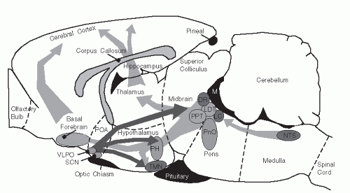
The involvement of the hypothalamus in sleep regulation was first suggested in the 1930s. At that time, a Viennese neurologist, Baron Constantin Von Economo, identified select populations of patients with symptoms of either prolonged insomnia or excessive sleepiness associated with endemic encephalitis. Upon autopsy, it was identified that those patients with prolonged insomnia had lesions located near the anterior hypothalamus in the region of the ventrolateral preoptic nucleus (VLPO). Conversely, patients with symptoms of excessive sleepiness were identified to have lesions of the posterior hypothalamus (PH). This led to the hypothesis that regions in the anterior and PH had opposing influences on the sleep-wake cycle. We discuss later how these hypothalamic regions interact with neurons of the brain stem reticular-activating system as well as histaminergic neurons of the tuberomammillary nucleus (TMN) and the SCN to promote sleep or wakefulness over a 24-hour cycle (Fig. 3-1).


Hypothalamus and Sleep


Suprachiasmatic Nucleus

Two general sleep control mechanisms are recognized: a homeostatic mechanism and a circadian mechanism. The homeostatic mechanism dictates that a given quota of sleep duration and intensity needs to be obtained over a short term and that current sleep needs depend on the individuals’ immediate prior history of sleep/wakefulness. Sleep deprivation causes a “rebound” effect in which, at the nearest available opportunity, an individual will sleep with an increased duration and intensity (increased cortical slow waves) to compensate for lost sleep (17,18 and 19). The circadian mechanism, located in the SCN of the hypothalamus, dictates that the animal should sleep during a set time frame each day. Both mechanisms are present in all species studied to date, including flies and scorpions. Yet how these two mechanisms interact to regulate an individual’s sleep cycle is not completely understood.

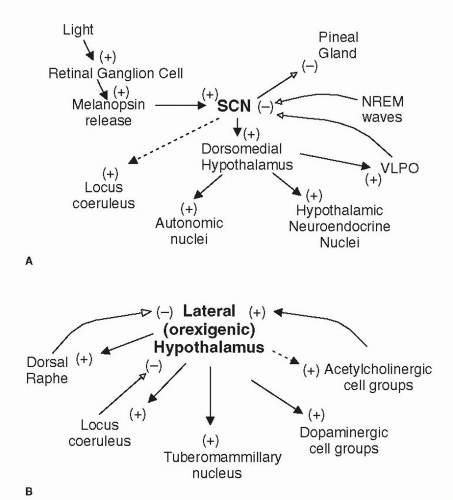
The SCN is thought to promote arousal by generating a signal that increases in strength throughout the biologic day (active period) and decreases at night. Loss of input from the SCN causes a loss of sleep consolidation (20,21). Pathways through which the circadian signal is transmitted to possible substrates of the homeostatic mechanism are being found (Fig. 3-2A). In rodents, the majority of SCN neurons project to the dorsomedial hypothalamus, which then acts as a distributor for SCN information. The dorsomedial hypothalamus sends projections to three major populations of target neurons (22). The first includes hypothalamic nuclei known to participate in sleep—wake regulation—the VLPO and the extended VLPO area. The second constitutes a group of hypothalamic neuroendocrine cells that secrete orexin, corticotrophin-releasing hormone, thyrotropin-releasing hormone, and gonadotrophin-releasing hormone (23). The third group constitutes autonomic cells that project to brain stem and spinal cord autonomic (both sympathetic and parasympathetic) nuclei. Apart from hypothalamic connections, an indirect connection through which the SCN
drives locus caeruleus (LC) neurons has been located (24). The SCN also influences melatonin and body temperature cycles directly, independent of dorsomedial hypothalamus contributions.


FIGURE 3-1 Schematic of location of sleep- and wake-promoting regions in the brain. Sagittal view of a rat brain redrawn from Paxinos and Watson’s The Rat Brain in Stereotaxic Coordinates (16) showing the location of major brain regions involved in wakefulness and sleep behavior. Monoaminergic nuclei are shown as dark-shaded circles. Cholinergic nuclei include the PPT and LDT nuclei. Other important nuclei are shown as light-shaded circles. Major ascending excitatory, wake-promoting projection pathways are illustrated by gray arrows, including the NTS, LC, LDT nucleus, PPT nucleus, DR, oral pontine region (PnO), the PH, including orexigenic neurons in the lateral hypothalamus, the TMN, and the SCN. The primary sleep-promoting site is located in the VLPO. The VLPO contains inhibitory neurons that project and suppress activity in wake-promoting regions.

Do the substrates for homeostatic control feedback information influence circadian rhythm generators? Sleep deprivation is known to disrupt the circadian rhythm (25). Sleep deprivation studies in rats show that the slow waves of NREM sleep feed back onto the circadian rhythm generators to slow the signal output from the generator (26). The faster waves of REM sleep increase the firing rate of SCN cells. Identities of the neural substrates, pathways, and transmitters are unknown, but evidence suggests that the SCN is able to track the amount of the different types of sleep and perhaps adjust its schedule accordingly.


Lateral Hypothalamus and Orexin

The lateral hypothalamus, located in the PH, is the exclusive source of recently discovered, arousal-promoting neuropeptides, orexin 1 and 2 (also known as hypocretin 1 and 2). Genetic deficiencies occurring in knockout mice (27) and human families indicate that signs of orexin deficiency include narcolepsy, cataplexy, decreased food intake, and perhaps metabolic disturbances (28).

Studies of orexin levels in the cerebrospinal fluid (CSF) have added information about its possible functions in normal individuals. Orexin levels in the CSF are lowest during NREM sleep and highest during wakefulness (29). This, together with findings that lateral hypothalamic neurons start firing before the transition from sleep to wakefulness, suggests a role for orexins in sleep—wake transitions (30). During wakefulness, levels are higher during activity than during quiet periods (31), causing many to believe that orexins are involved in promoting motor, especially food-seeking activity

Orexin-producing lateral hypothalamic neurons send abundant, strong, excitatory projections to wakepromoting noradrenergic, histaminergic, dopaminergic, and cholinergic nuclei (32) and are likely to regulate these centers (Fig. 3-2B). In turn, slice studies show inhibitory effects of noradrenaline and serotonin on orexin production, whereas acetylcholine input excites orexin-producing cells (33). This arrangement suggests the presence of self-regulating feedback loops. In addition, the SCN influences orexin production according to the individuals’ circadian rhythm.

On the basis of information at hand, Siegel (32) has hypothesized that the role of orexins is “to facilitate motor activity in association with motivated behaviors and coordinate this with activation of attentional and sensory systems.”


Ventrolateral Preoptic Nucleus

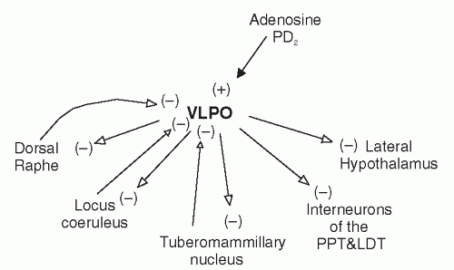
Opposing the arousing effect of posterior hypothalamic and brain stem centers is a “sleep-generating” nucleus in the anterior hypothalamus, in the preoptic area. Immunostaining with c-Fos has identified two groups of sleepactive neurons in this area (34). The first, located in the VLPO, is associated with NREM sleep (35). The second, located dorsal and medial to the first (the extended VLPO), is more closely linked to REM sleep (36). VLPO neurons are activated by sleep-inducing factors, such as adenosine and prostaglandin D2 (37,38) (Fig. 3-3). They are also temperature (warm) sensitive (39) and so compatible with the effect of temperature on the propensity for sleep.


FIGURE 3-2 Interconnections of wake-promoting brain regions. (A) Influence of SCN on sleep-associated centers, autonomic, and hormonal control centers. (B) Influence of lateral hypothalamus on wake-promoting regions in the brain stem and forebrain. Solid lines indicate strong interconnections. Dashed lines indicate weak interconnections. Filled arrows and (+) represent excitatory connections. Open arrows and (-) represent inhibitory connections.

Neurons in the VLPO contain the inhibitory transmitters, γ-aminobutyric acid (GABA) and galanin (40,41), and project to locations of “arousal” neurons, including the serotonergic dorsal raphe (DR) cell group, histaminergic TMN, noradrenergic LC, and the orexigenic lateral hypothalamic nucleus (40,42,43). The VLPO also projects to cholinergic nuclei (basal forebrain and pedunculopontine tegmental [PPT] nucleus and laterodorsal tegmental [LDT] nucleus) but primarily innervates inhibitory interneurons within these regions. Projections to LC, raphe, and PPT—LDT are derived mostly from the extended VLPO, rather than the VLPO (44). In return, VLPO neurons receive inhibitory adrenergic and serotonergic input from the LC and raphe nuclei, respectively (45,46). GABA release into arousal areas increases during NREM sleep (47), and the VLPO has been shown to regulate the amount of delta activity, an index of sleep intensity, within NREM sleep. Because activation of VLPO neurons appears to be required for normal regulation of sleep, the VLPO is considered an essential element of the sleep-wake central circuitry.


Tuberomammillary Nucleus

It has long been recognized that antihistamines have a powerful sedative action, yet the presence of histaminergic neurons in the brain was not identified until 1984, when appropriate immunohistochemical techniques became available (48,49). To date, histaminergic neurons have been found localized to the PH in the region of the TMN of the hypothalamus. Histaminergic neurons in the TMN project throughout the central nervous system (CNS), including the cerebral cortex, amygdala, and substantia nigra, three regions that receive the densest innervation. The TMN receives input from orexigenic neurons in the lateral hypothalamus and has descending projections to sleep-related regions of the brain stem, including LC, DR,
the parabrachial nucleus, and nucleus of the solitary tract (NTS) in the dorsal medulla. Many brain stem reticular formation regions, in turn, send ascending projections back to the TMN (Fig. 3-1). The LC and ventral tegmental regions, however, send few fibers to the TMN. TMN neurons also receive afferent input from the GABAergic neurons in the VLPO, which appear to contribute strongly to the firing rate of these histaminergic neurons in relation to behavioral state.


FIGURE 3-3 Interconnections of sleep-promoting brain regions. Projection sites of the inhibitory neurons in the VLPO are important in promoting sleep. VLPO are activated by wake-promoting influences such as adenosine and prostaglandin D2. Filled arrows and (+) represent excitatory connections. Open arrows and (-) represent inhibitory connections.

Histaminergic neurons in the TMN have pacemakerlike activity and fire with characteristically high rates during wakefulness, slower rates during NREM sleep, and almost completely cease discharging during REM sleep, similar to the pattern observed in the monoaminergic neurons in the brain stem reticular-activating system.


Pineal Gland

The pineal gland is positioned on the posterodorsal part of the third ventricle and, typical of all endocrine glands, it is well vascularized. The pineal gland secretes melatonin into the surrounding cerebral sinuses in response to photic information received primarily by the eyes (Fig. 3-1). Melatonin is best known for its ability to induce seasonal and circannual physiologic changes in animal species, for example, the reproductive propensity in sheep. Melatonin levels also fluctuate on a daily basis according to the individuals’ light exposure, yet its influence on the circadian rhythm and sleep—wake cycle is less clear.

Retinal exposure to light, especially in the range of 460 to 470 nm (50), inhibits melatonin release. Upon exposure to light, retinal ganglion cells release a recently discovered molecule, melanopsin, into the SCN (51). The SCN response signal passes through the sympathetic intermediolateral cell column in the thoracic spinal cord and returns to the pineal gland to inhibit melatonin release. Melatonin suppression can still occur in some blind people since its secretion is independent of the integrity of rods and cones. However, if retinal ganglion cells are also damaged, melatonin fluctuations may become independent of light exposure. Because of its direct and strong link to the SCN, and because it is relatively easily measured in saliva, melatonin is often used as a marker of circadian rhythm (52).

The effects of melatonin on physiologic rhythms can be established by studying pinealectomized animals. Pinealectomy eliminates the response to changing daylight lengths in several species, and suitable melatonin replacement restores the seasonal cycles. However, the effects on the daily cycle are subtle. Rats adapt faster to light—dark phase shifts in the absence of a pineal gland (52). Reports of humans with a pinealectomy are rare, and consistent consequences of lacking the gland have been unable to be established.

Only gold members can continue reading. Log In or Register to continue

Stay updated, free articles. Join our Telegram channel

Jul 14, 2016 | Posted by in PSYCHIATRY | Comments Off on The Neurobiology of Sleep

Full access? Get Clinical Tree

Get Clinical Tree app for offline access