The neurological examination: Peripheral nervous system

4 The neurological examination


Peripheral nervous system


Examination of the peripheral nervous system assumes a stylised approach when adopting the traditional method. This means following a set pattern of: observation and inspection; tone; power; reflexes; sensation; coordination; and gait. This ignores what was stated earlier, namely that the consultation starts long before the patient enters the consultation room.


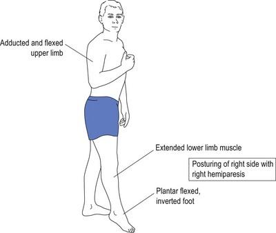
The first part of testing of the peripheral nervous system is observation, namely observing how the patient gets out of the chair and walks into the room. Difficulty rising from the chair may suggest Parkinson’s disease. Not swinging one arm when walking may suggest either pyramidal disease, such as a stroke, or may also suggest Parkinson’s disease. Spastic posturing (flexed upper limb and extended lower limb) with circumduction of affected lower limb suggests upper motor neurone damage, such as stroke. Stooped posture, shuffling gait with festination, hesitancy and inertia of gait initiation are almost diagnostic of Parkinson’s disease, and are apparent long before the consultation formally commences.


Ataxic, wide-based gait is suggestive of cerebellar disease. Wide-based gait is far less common than one would think. The width between the feet is usually wider than the shoulders of the patient and gives the impression that the patient is drunk—hence the statement that the patient walks like a drunken sailor. Wide-based gait provides stability of stance, and sailors used to moving ships often walk with such wide-based gait to maintain stability on a ship.


Before undergoing the formal peripheral assessment it is worth testing for grasp reflexes. This is done by distracting the patient, possibly by engaging in ‘small talk’, and while doing so sliding the hand out, pushing up against the patient’s palm and fingers. Grasp reflex may be as subtle as feeling the fingers of the patient flexing downwards towards the examiner’s sliding fingers. A positive grasp reflex is indicative of contralateral frontal lobe (upper motor neurone) damage. Other frontal lobe signs may include palmar–mental response. This is evoked by applying a noxious stimulus (such as scratching) to the palm of the patient’s hand and observing the movement of the chin (mentalis muscle) on the same side as the scratched hand. This again suggests contralateral frontal lobe damage.


The glabellar tap is elicited by tapping the index finger on the patient’s forehead. It is best achieved by holding the hand above the forehead and tapping the forehead without coming front-on as this movement, equivocal to menace (as described when testing visual fields in Ch 2), may itself evoke a blink response. When testing the glabellar tap the normal response allows up to three blinks. More than three blinks represent a positive response. When testing glabellar tap it is worth reappraising the patient for ptosis, as attention is focused on the eyelids.


Much of the above actually tests central nervous system function, but it is best examined at this point in patient assessment. This uses the peripheral to test central competence.


While the approach adopted has been compartmentalised, this has been done for ease of understanding. An additional consideration has been to instil time-efficient consultation methods: all busy doctors are ‘time poor’ and anything that can improve clinical acumen and save time is of value.


Tone


Tone is tested by a number of different methods. Initial resistance, followed by a feeling of giving way, is called ‘clasp knife’ as it is similar to opening the blade of a pocketknife. This is found with spasticity in upper motor neurone damage.


If Parkinson’s disease is considered, then the initial testing of tone by moving the hand up and down at the wrist should be normal early in the disease process. The patient is asked to move their head from side to side, pushing the ear into the shoulder with each turn, at which time the tone increases with ‘cogwheeling’ and ‘lead pipe’ rigidity. This is pathognomic of early Parkinson’s disease.


If the patient is examined soon after the onset of a stroke, the hemiplegic limbs may be totally flaccid and floppy. After longstanding upper motor neurone damage, the hyper-tonicity may progress to clonus. This is evoked by short, sharp joint movement, such as sudden sharp dorsiflexion of the foot, similar to the initial provocative jerking movement. The same may be caused by jerking the patella bone towards the foot in the extended knee. This may evoke repeated movement of the knee bone up and down in a similar fashion to that seen at the foot. Clonus may be evoked at other joints, such as the wrist, but lower limb clonus is more common.


Cerebellar damage does not cause increased tone which is often decreased, causing pendular movements on testing. This is often difficult for the inexperienced to discern. While increased or decreased tone is relatively easy to identify, often the subtleties of tone are not that easy to identify for the doctor not attuned to the neurological examination.


Power


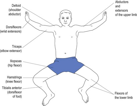
Various conditions affect different muscles. Upper motor neurone weakness, as with stroke, affects the antigravity muscles. Most inexperienced doctors have difficulty remembering which muscles constitute the antigravity muscles (see Fig 4.1).


It is because of weakness of the antigravity muscles that the patient with upper motor neurone damage presents with flexion of the upper limbs and extension of the lower limbs. With weakness of the antigravity muscles, the opposing muscles exert their effect causing posturing without counter effect by opposing muscles (see Fig 4.2).


To make it simpler for the doctor, it is easier to remember just one group of muscles, such as the extensors in the upper limbs. It is even easier to remember just one upper limb muscle, such as the triceps, which is an extensor muscle. This will reinforce extensors in the upper limbs and the opposite, namely the flexors with lower limbs. Alternatively, the doctor may choose to remember just a single flexor in the lower limb, such as the hip flexor, the iliopsoas.


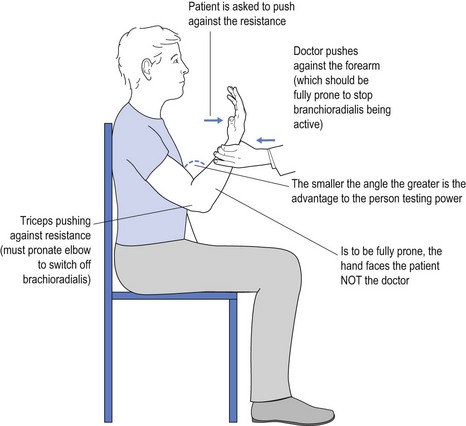
It is obvious that a 60 kg female may have trouble overcoming the strength of a 100 kg male who is engaged in very physical work. One way to overcome this is to use mechanical advantage to test power. An example of this might be to test triceps power by flexing the elbow much more than the traditional 90° (see Fig 4.3).


< div class='tao-gold-member'>

Stay updated, free articles. Join our Telegram channel

Jun 19, 2016 | Posted by in NEUROLOGY | Comments Off on The neurological examination: Peripheral nervous system

Full access? Get Clinical Tree

Get Clinical Tree app for offline access