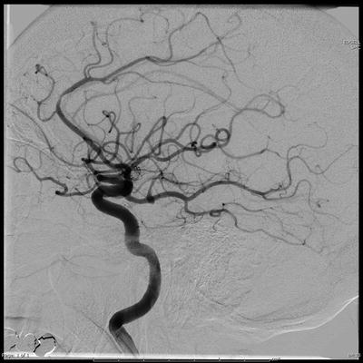
Fig. 9.1
Lateral projection image of a persistent trigeminal artery. These fetal remnants may allow intra-arterial injections to affect the brainstem and thalami, potentially leading to impairment of consciousness and respiratory depression
A standard anterior–posterior (AP) and lateral cerebral angiogram is performed on both sides to identify any relevant anatomical variations in the cervical or cranial vasculature. Angiography of the posterior circulation is not performed routinely, except when needed to further define unusual vascular anatomy in the carotid distribution or if selective PCA barbiturate injections will be performed.
Special Anatomical Considerations (See Chap. 2)
Fetal PCA
Patients with a fetal-type PCA may have filling of the basilar artery during routine angiographic injections (Fig. 9.2). In these patients, a rapid infusion of barbiturates may result in respiratory suppression, lethargy, and even autonomic instability due to brainstem compromise. Wada testing can be performed safely in patients with a fetal PCA provided the barbiturates are injected slowly, as competitive flow from the basilar artery will usually prevent the drug from flowing into the posterior circulation. This can be confirmed by doing a control run using contrast prior to drug infusion. In the rare case where a slow injection is not sufficient to prevent reflux into the basilar artery, the drug can be injected directly into the MCA using a microcatheter.


Fig. 9.2
Lateral projection image of a fetal posterior cerebral artery. This common anatomical variant does not preclude carotid injection, but care must be taken to administer the agent gradually in order to avoid filling of the posterior circulation
Persistent Carotid–Basilar Anastomosis
Persistent carotid–basilar (PCB) anastomoses are an uncommon anatomical variant of the circle of Willis in which there is a direct connection between the carotid artery and the vertebrobasilar system (Fig. 9.1). Unlike the more common fetal PCA variant, patients with a PCB anastomosis often will have unavoidable filling of most if not the entire basilar artery during a routine cervical carotid contrast injections. Consequently, cervical carotid injections of barbiturates during Wada testing may result in severe respiratory depression, lethargy, and even autonomic instability. Wada testing can be safely performed in patients with a PCB anastomosis if the barbiturates are injected into the carotid artery distal to the anastomosis using a microcatheter or balloon microcatheter.
Absent or Atretic A1
Patients with an absent or atretic ACA artery may have nearly all of the blood to the frontal lobes supplied by one carotid artery. Injections on the side of the dominant ACA will often cause bilateral frontal lobe suppression leading to behavioral changes such as dysinhibition and confusion. This effect may be particularly profound when the dominant A1 is on the right side. In patients with an atretic but functional contralateral ACA, a softer injection will help to prevent the reflux of barbiturates into the contralateral hemisphere. In patients with absence of the contralateral ACA, direct injection of barbiturates into the MCA with a microcatheter on the side of the dominant A1 can be considered.
Carotid Occlusion
Rarely, a patient will be found to have an asymptomatic carotid artery occlusion or high-grade stenosis. In cases where testing from the ipsilateral carotid is not possible, barbiturate injections can be performed from the PCAs using a microcatheter.
Intracarotid Barbiturate Injections
A diagnostic cerebral angiogram is performed in the hemisphere giving rise to seizures. If there are no complicated vascular anatomical features, the catheter is then flushed with heparinized saline, and the C-arm is repositioned to allow the neurologist and neuropsychologist free access to the patient’s head and arms. The patient is then given a brief tutorial regarding the testing procedure.
The test is initiated by having the patient raise both arms (braced by the neurology team to avoid contamination of the sterile field). As the patient counts backward from 100 aloud, a bolus of the anesthetic of choice is administered through the diagnostic catheter until the patient’s contralateral upper extremity becomes flaccid. Once the neurologist determines the bolus was effective, the neuropsychological testing is performed. During neuropsychological testing, the neurologist continuously assesses the efficacy of the anesthetic dose by looking for the return of muscle tone or movement in the affected extremity and by inspecting the EEG pattern. Once the neuropsychological testing is complete, the catheter is removed from the body and flushed. After the patient recovers from the effects of the anesthetic, the second phase of neuropsychological testing of memory is completed along with assessment to ensure the patient has not had a stroke. The process is then repeated on the other side.
Potential Pitfalls
The Wada test has all the inherent risks associated with diagnostic cerebral angiography including arterial dissection, stroke, and complications from bleeding but also has some unique consequences that may not be encountered in routine diagnostic testing.
Embolus
Emboli during Wada testing can occur as the result of clot formation on wires or catheters used for access, by dislodging plaque in the carotid artery or arch, or due to introduction of foreign particulate matter during contrast or drug injections. When an embolus is observed, whether symptomatic or not, the test must be discontinued. The catheter should be bled back and flushed to remove any particulate material. For small, distal emboli that cannot be reached with a clot retriever, consider administration of IA abciximab (0.25 mg/kg) through the arterial catheter or IA tPA (1–2 mg). A retrospective study of 677 patients showed that the incidence of stroke and the incidence of transient ischemic attack were both 0.6 % [3].
Seizure
A complex partial or generalized tonic–clonic seizure during Wada testing is a rare but not completely unexpected event with an incidence of 1.2 % [3]. Supportive care includes removing the diagnostic catheter and turning the head to the side to avoid aspiration. The use of lorazepam (2–4 mg) depends upon the patient’s history of isolated or serial seizures. Since the postictal state following a seizure may influence the results of neuropsychological testing, the Wada is typically discontinued.
Pediatric Patient Testing
Wada testing of children 13 years and older is generally well tolerated without special accommodations. Testing of preteen children as young as 6 years has been shown to be safe and effective provided appropriate pre-procedure training is performed. Mild sedation with propofol during femoral access and control angiography in these young patients have been used to help improve comfort and compliance during Wada testing [4–6].
Pharmacology
Methohexital (Brevital)
Methohexital (JHP Pharmaceuticals, Parsippany, NJ) is supplied as a lyophilized powder. The powder is reconstituted in sterile water or normal saline to a concentration of 10 mg/ml. It is then passed through a syringe filter onto the sterile field where it is further diluted with preservative-free saline to a final concentration of 1 mg/ml. An amount of 10 ml of methohexital (1 mg/ml) is drawn into a 10 ml syringe in preparation for injection.
The onset of action is nearly immediate (2–3 s). In most cases, 3–4 mg of methohexital is needed to initiate anesthesia. Boluses of 1–2 mg of methohexital every 90–120 s will be needed to maintain anesthesia during neurological testing. Full clinical and EEG recovery will take around 5–7 min [7].
Amobarbital (Amytal)
Amobarbital (Marathon Pharmaceuticals, Deerfield, IL) is supplied as a lyophilized powder. It is traditionally the first-line agent for these procedures but has been subject to supply shortages. The drug is diluted in 5 ml of sterile water and is dissolved by rotating the vial—not shaking. It may take several minutes to completely dissolve. Additional saline is added to achieve a final concentration of 25 mg/ml. The final solution is then passed through a syringe filter on the field to remove any particulate matter. Amobarbital should be used within 30 min of preparation.
Amytal dosing in adults is 125 mg, 5 ml administered at 1–2 ml/min. Up to an additional 50 mg (1–2 ml) may be administered, if needed, to induce hemiparesis. Lower doses of amobarbital (75 mg) have also been used effectively in adults in experienced centers, despite conflicting results from historical reports [8].
Etomidate (Amidate)
Etomidate (Hospira, Lake Forest, IL) is a nonbarbiturate hypnotic drug with a rapid onset of action. The liver rapidly metabolizes it. Etomidate is supplied as a sterile solution at a concentration of 2 mg/ml and can be diluted with saline. The onset of action after IA injection is approximately 45 s to 1 min, and its effects last approximately 5 min [9].
Stay updated, free articles. Join our Telegram channel

Full access? Get Clinical Tree


