FIGURE 19.1 Relationship of the cranial and spinal portions of the accessory nerve to the vagus and glossopharyngeal nerves.
The major part of CN XI is the spinal portion (ramus externus). Its function is to innervate the sternocleidomastoid (SCM) and trapezius muscles. The fibers of the spinal root arise from SVE motor cells in the SA nuclei in the ventral horn from C2 to C5, or even C6. The cell column of the SA nucleus lies in a position analogous to the nucleus ambiguus in the medulla. The cell column making up the SA nucleus is somatotopically organized. The upper spinal cord portion innervates primarily the ipsilateral SCM; the lower spinal portion innervates primarily the ipsilateral trapezius. In keeping with the tendency of branchial arch–related nerves to make internal loops, its axons arch posterolaterally through the lateral funiculus, and emerge as a series of rootlets laterally between the anterior and posterior roots. These unite into a single trunk, which ascends between the denticulate ligaments and the posterior roots. The nerve enters the skull through the foramen magnum, ascends the clivus for a short distance, and then curves laterally. The spinal root joins the cranial root for a short distance, probably receiving one or two filaments from it. It exits through the jugular foramen in company with CNs IX and X.
CN XI emerges from the skull posteromedial to the styloid process and then descends in the neck near the internal jugular vein, behind the digastric and stylohyoid muscles, to enter the deep surface of the upper part of the SCM muscle. It passes through the SCM, sending filaments to it, and then emerges at its posterior border near the midpoint, coursing near the great auricular nerve. CN XI then runs obliquely across the posterior triangle of the neck on the surface of the levator scapula muscle, superficially and in close proximity to the lymph nodes of the posterior cervical triangle. About three fingerbreadths above the clavicle, the nerve enters the deep surface of the anterior border of the upper trapezius muscle. In the neck, the SA contributes fibers to the cervical plexus, rami trapezii, and then courses to the caudal aspect of the trapezius. Most of the communications with C2 through C4 are conveying proprioceptive information from CN XI, which will enter the spinal cord in the upper cervical segments. The innervation of the SCM may be more complex than is to be found in most anatomical texts, possibly including fibers from CN X. Over half of the patients undergoing division of the SA nerve and the upper cervical motor roots as treatment for cervical dystonia had residual SCM activity of sufficient magnitude to make further surgery necessary before the muscle was effectively paralyzed.
The innervation of the trapezius shows some individual variability. The SA nerve is the primary innervation to the upper trapezius, but Soo et al. have shown that the cervical plexus may contribute motor fibers, especially to the middle and lower trapezius. The neurons of the spinal portion of XI communicate with the oculomotor, trochlear, abducens, and vestibular nuclei through the medial longitudinal fasciculus. These connections are important in controlling conjugate deviation of the head and eyes in response to auditory, vestibular, and other stimuli.
The supranuclear innervation of CN XI arises from the lower portion of the precentral gyrus. Fibers from the lateral corticospinal tract in the cervical spinal cord communicate with the SA nucleus. There is some controversy, but the bulk of current evidence indicates that both the SCM and trapezius receive bilateral supranuclear innervation. However, the input to the SCM motor neuron pool is predominantly ipsilateral and that to the trapezius motor neuron pool is predominantly contralateral (Box 19.1). The SCM turns the head to the opposite side, and its supranuclear innervation is ipsilateral. Therefore, the right cerebral hemisphere turns the head to the left.
Cortical Innervation of the Sternomastoid Muscle
The sternocleidomastoid (SCM) may be an exception to the general scheme of contralateral hemispheric innervation. Authorities debate whether the SCMs receive ipsilateral, contralateral, or bilateral cortical innervation. Studying the function of the SCM muscles during intracarotid injection of amytal (Wada testing), DeToledo et al. demonstrated weakness of the right SCM after injection into the right internal carotid artery in some patients and little to no weakness in others. This suggested that the SCMs receive bilateral hemispheric innervation with the maximal input from the ipsilateral hemisphere. They proposed that the SA nucleus has rostral and caudal portions and that the rostral (SCM) portion receives bihemispheric projections, but the innervation of the caudal (trapezius) portion is predominantly contralateral. This would be analogous to the supranuclear innervation of the facial nerve nucleus, bilateral to the rostral portions but contralateral to the caudal portions. Transcranial stimulation studies concluded projections to the SCM were bilateral but predominantly contralateral, and those to the trapezius exclusively contralateral. Some have contended the nerve double decussates, but this remains unproven.
The SCM muscles function with the other cervical muscles to flex the head and turn it from side to side. When one SCM contracts, the head is drawn toward the ipsilateral shoulder and rotates so that the occiput is pulled toward the ipsilateral shoulder, while the face turns in the opposite direction and upward. Acting together, the two muscles flex the neck and bring the head forward and downward. With the head fixed, the two muscles assist in elevating the thorax in forced inspiration.
The trapezius retracts the head and draws it ipsilaterally. It also elevates, retracts, and rotates the scapula and assists in abducting the arm above horizontal. When one trapezius contracts with the shoulder fixed, the head is drawn to that side. When both contract, the head is drawn backward and the face deviated upward. When the head is fixed, the upper and middle fibers of the trapezius elevate, rotate, and retract the scapula and shorten the distance between the occiput and the acromion. The lower fibers depress the scapula and draw it toward the midline. The SCM and trapezius muscles thus act together to rotate the head from side to side and to flex and extend the neck.
CLINICAL EXAMINATION
The functions of the cranial portion of CN XI cannot be distinguished from those of CN X, and examination is limited to evaluation of the functions of the spinal portion. A complex array of many muscles is involved in moving the head, including the scaleni, splenii and obliqui capitis, recti capitis, and longi capitis and colli. With bilateral paralysis of CN XI innervated muscles, there is diminished but not absent neck rotation, and the head may droop or even fall backward or forward, depending upon whether the SCMs or the trapezii are more involved.
One SCM acts to turn the head to the opposite side or to tilt it to the same side. Acting together, the SCMs thrust the head forward and flex the neck. The muscles should be inspected and palpated to determine their tone and volume. The contours are distinct even at rest. With a nuclear or infranuclear lesion, there may be atrophy or fasciculations.
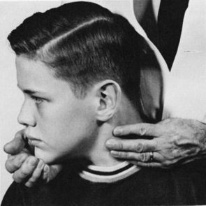
To assess SCM power, have the patient turn the head fully to one side and hold it there, then try to turn the head back to midline, avoiding any tilting or leaning motion. The muscle usually stands out well, and its contraction can be seen and felt (Figure 19.2). Significant weakness of rotation can be detected if the patient tries to counteract firm resistance. Unilateral SCM paresis causes little change in the resting position of the head. Even with complete paralysis, other cervical muscles can perform some degree of rotation and flexion; only occasionally is there a noticeable head turn. The two SCM muscles can be examined simultaneously by having the patient flex his neck while the examiner exerts pressure on the forehead or by having the patient turn the head from side to side. Flexion of the head against resistance may cause deviation of the head toward the paralyzed side. With unilateral paralysis, the involved muscle is flat and does not contract or become tense when attempting to turn the head contralaterally or to flex the neck against resistance. Weakness of both SCMs causes difficulty in anteroflexion of the neck, and the head may assume an extended position. The SCM reflex may be elicited by tapping the muscle at its clavicular origin. Usually, there is a prompt contraction. The reflex is mediated by the accessory and upper cervical nerves, but has little significance in neurologic diagnosis.

FIGURE 19.2 Examination of the sternocleidomastoid (SCM) muscle. When the patient turns his head to the right against resistance, the contracting muscle can be seen and palpated.
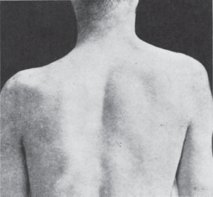
With trapezius atrophy, the outline of the neck changes, with depression or drooping of the shoulder contour and flattening of the trapezius ridge (Figure 19.3). Severe trapezius weakness causes sagging of the shoulder, and the resting position of the scapula shifts downward. The upper portion of the scapula tends to fall laterally, while the inferior angle moves inward. This scapular rotation and displacement are more obvious with arm abduction.

FIGURE 19.3 Paralysis of the left trapezius muscle. There is a depression in the shoulder contour with downward and lateral displacement of the scapula.


