The Vestibular System: Introduction
Anatomy
The membranous labyrinth, filled with endolymph and surrounded by perilymph, lies in the bony labyrinthine space within the temporal bone of the skull base (Fig 17–1). Two special sensory systems receive their input from structures in the membranous labyrinth: the auditory system, from the cochlea (see Chapter 16), and the vestibular system, from the remainder of the labyrinth.
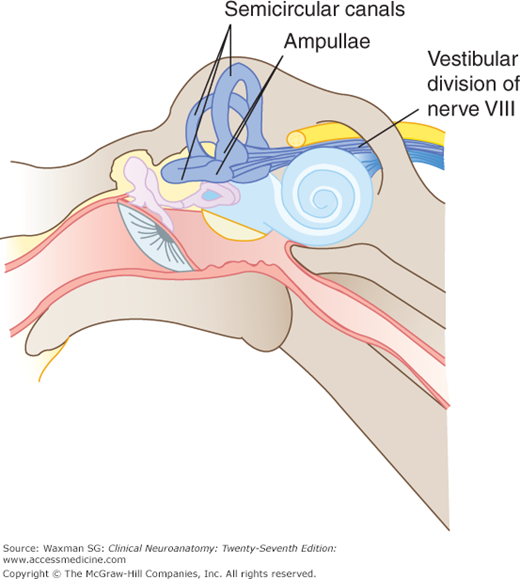
The static labyrinth gives information regarding the position of the head in space; it includes the specialized sensory areas located within the saccule and the utricle (see Fig 17–1). Within the utricle and saccule, otoliths (small calcium carbonate crystals, also termed otoconia) are located adjacent to hair cells clustered in macular regions. The otoliths displace the hair cell processes and excite the utricle and saccule in response to horizontal and vertical acceleration.
The kinetic labyrinth consists of the three semicircular canals. Each canal ends in an enlarged ampulla, which contains hair cells, within a receptor area called the crista ampullaris. A gelatinous partition (cupula) covers each ampulla and is displaced by rotation of the head, displacing hair cells so that they generate impulses. The three semicircular canals are oriented at 90°to each other, providing a mechanism that is sensitive to rotation along any axis.
Vestibular Pathways
The peripheral branches of the bipolar cells in the vestibular ganglion course from the specialized receptors (hair cells) in the ampullae and from the maculae of the utricle and the saccule. The central branches run within the vestibular component of cranial nerve VIII to enter the brain stem and end in the vestibular nuclei (Figs 17–1 and 17–2; see also Chapter 7).
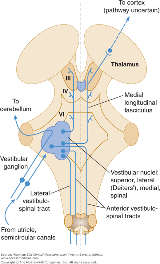
Some vestibular connections go from the superior and lateral vestibular nuclei to the cerebellum, where they end in the cerebellar cortex within the flocculonodular component (see Chapter 7). Others course from the lateral vestibular nuclei into the ipsilateral spinal cord via the lateral vestibulospinal tracts, from the superior and medial vestibular nuclei to nuclei of the eye muscles, and to the motor nuclei of the upper spinal nerves via the medial longitudinal fasciculi (MLF) of the same and opposite sides (Fig 17–2). The medial vestibulospinal tract (the descending portion of the MLF) connects to the anterior horn of the cervical and upper thoracic cord; this tract is involved in the labyrinthine righting reflexes that adjust the position of the head in response to signals of vestibular origin. Some vestibular nuclei send fibers to the reticular formation. Some ascending fibers from the vestibular nuclei travel by way of the thalamus (ventral posterior nucleus) to the parietal cortex (area 40).


