Theta Activity
▲ Cigánek Rhythm
Other Names
Episodic anterior drowsy theta
Frontal midline theta rhythm
Midline theta rhythm
Description
Descriptions of the simply termed Cigánek rhythm are highly consistent with regard to frequency, frequency range, location, distribution, and waveform but less in agreement about duration. A Cigánek rhythm’s frequency is almost always between 5 and 7 Hz and most often is 6 Hz (Niedermeyer, 1999b; Westmoreland, 1990). The field is centered at the midline with a maximum that usually is near the vertex (Cz) and may be displaced by one electrode distance anteriorly (Fz) or posteriorly (Pz); however, an anterior displacement occurs much more commonly than a posterior displacement. The field’s distribution may or may not include the immediately parasagittal electrodes. The waveform includes a rising and falling amplitude over the pattern’s occurrence with a maximum amplitude that usually is greater than 50 μV and wave elements that usually are sinusoidal but may be arciform. Other than the rising and falling amplitude, the rhythm does not evolve. Although some reports include a duration definition of greater than 3 seconds and as long as 20 seconds, others have described similar rhythms lasting less than 3 seconds (Janati et al., 1986; Okada and Urakami, 1993; Westmoreland and Klass, 1986).
Distinguishing Features
• Compared to Alpha Activity’s mu Rhythm
The central location and occasional arciform appearance are the overlapping features of the mu and Cigánek rhythms. However, these two rhythms are easily distinguishable by frequency and field. The mu rhythm is within the alpha frequency range and is usually asymmetrically placed in the parasagittal region. The mu rhythm characteristically attenuates with upper extremity movement, but this is not always reliable as a distinguishing feature because movement may be accompanied by arousal, which can cause the Cigánek rhythm to attenuate (Westmoreland and Klass, 1986).
• Compared to Hypnopompic, Hypnagogic, and Hedonic Hypersynchrony
The hypersynchronous patterns and the Cigánek rhythm can have similar rhythmic theta activity, but the field for hypersynchronous patterns is substantially broader and the amplitude is usually higher. Hypersynchronous patterns span the central region and extend to be similar to the generalized patterns in their distribution. The greater amplitude of hypersynchronous patterns produces the appearance of a more abrupt development and greater differentiation from the background activity than the Cigánek rhythm. The duration is not as reliable as a distinguishing feature, but hypersynchronous patterns often last much longer than typical Cigánek rhythms.
• Compared to Polymorphic Theta Activity
Polymorphic theta frequency range activity may accompany the Cigánek rhythm because both patterns occur during early stages of sleep, although the Cigánek rhythm more typically occurs before sleep onset and polymorphic theta activity usually is most pronounced after sleep onset. However, the two patterns are more readily distinguished through their waveforms, durations, and distributions. Unlike the Cigánek rhythm, polymorphic theta activity is not a discrete regular rhythm. It includes several frequencies within the theta frequency range, and, partly because of the mixture of frequencies, it does not have as clear a beginning and end. Polymorphic theta activity also extends beyond the central region into a more generalized or lateralized distribution.
• Compared to Saw-Tooth Waves of Rapid Eye Movement Sleep
Saw-tooth waves of rapid eye movement (REM) sleep share location and sometimes frequency with the Cigánek rhythm; however, saw-tooth waves usually have a frequency in the delta frequency range. When occurring in the theta frequency range, saw-tooth waves can be distinguished by their waveform and the behavioral state in which they occur. Saw-tooth waves have a sharp contour that gives the pattern its descriptive name. The Cigánek may be sharply contoured when it has an arciform appearance, but an arciform contour differs from a saw-tooth waveform because of the combination of a rounded phase and pointed phase. Furthermore, the Cigánek rhythm occurs in drowsiness and saw-tooth waves of REM sleep are specific for REM sleep; thus, the accompanying waves are helpful in distinguishing the patterns. REM sleep is accompanied by minimal movement and muscle artifact, rapid eye movement artifact, and a mixture of frequencies, including activity in the alpha frequency range. The drowsy and light sleep states typical for a Cigánek rhythm have occasional movement or muscle artifact, slow eye movement artifact, and slower EEG background frequencies.
Co-Occurring Pat terns
Drowsiness or light sleep is the state in which a Cigánek rhythm most often occurs, but it has been described to also sometimes accompany concentration and it may be related to working memory (Inanaga, 1998; Sauseng et al., 2010). When occurring in drowsiness, the Cigánek rhythm may be accompanied by generalized, polymorphic theta activity, occasional slower waves, frontal–central beta activity, vertex sharp transients, and the absence of an alpha rhythm. It does not occur deeper than stage 1 of NREM sleep, so it is not accompanied by sleep spindles, K complexes, or a background of predominantly delta frequency range activity (Janati et al., 1986). Its occurrence with concentration is accompanied by other signs of wakeful attentiveness, including eye blink artifact and an intermittent alpha rhythm.
Clinical Significance
Although Cigánek’s original description of this rhythm in 1961 included the observation that it is abnormal and associated with epilepsy, subsequent publications have conclusively demonstrated that it is a normal finding in drowsiness or cognitive tasks (Ciganek, 1961; Westmoreland and Klass, 1986). However, its occurrence in relaxed wakefulness has been reported to be more common in frontal lobe epilepsy than in temporal lobe epilepsy or controls, and especially more common in medial frontal lobe epilepsy (Beleza et al., 2009). It also has been observed to be associated with euphoric states related to intoxication or exercise (Matsuoka, 1990).
The reported prevalence of this rhythm varies with how strictly it is defined and whether the population is one of patients or healthy controls. Overall, it is usually close to 1% for patients and about 6% for healthy controls (Okada and Urakami, 1993; Shinomiya et al., 1994). Based on multiple reports, its prevalence appears to peak in early adulthood, but it has only a minor decrease during adulthood into late life.
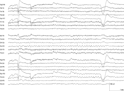 Figure 29-1 Cigánek Rhythm • The 6-Hz rhythm that is maximal at the Cz electrode is typical for a Cigánek rhythm in its frequency and location. Its 2-second duration, slight asymmetry with left-sided predominance, and somewhat arciform morphology are acceptable Cigánek rhythm variants. The rhythm is accompanied by frequent eye blink artifact and sustained EMG artifact, both of which may co-occur. (LFF 1 Hz, HFF 70 Hz) |
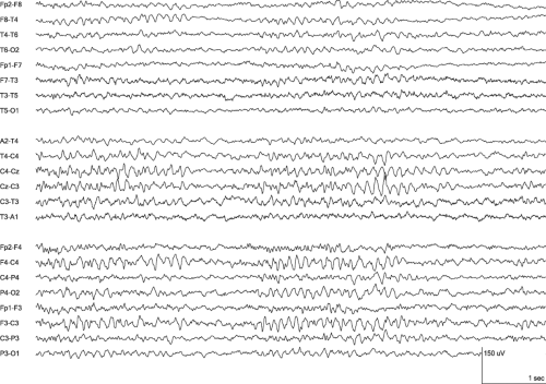 Figure 29-2 Cigánek Rhythm • Compared to Figure 29-1, the Cigánek rhythm in this segment is more symmetric, has a greater amount of intermixed faster frequencies, and occurs within slower background activity. All of these features make the Cigánek rhythm less apparent. Drowsiness is evident in the central beta activity and slow roving eye movement artifact. (LFF 1 Hz, HFF 70 Hz) |
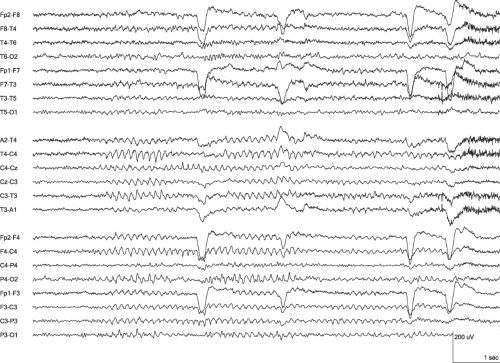 Figure 29-3 Cigánek Rhythm • The 7-Hz Cigánek rhythm is sustained through the first three occurrences of eye blink artifact and then ends with the fourth eye blink and increase in muscle artifact. The combination of eye blink and increased muscle artifact indicates an increase in wakefulness, which is a state change that can diminish a Cigánek rhythm. (LFF 1 Hz, HFF 70 Hz) |
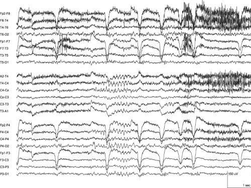 Figure 29-4 Cigánek Rhythm • The Cigánek rhythm lasts approximately 1 second and occurs amid frequent eye blink artifact and sustained and varying muscle artifact. The occurrence of a Cigánek rhythm in this state is consistent with its association with cognitive tasks requiring concentration. (LFF 1 Hz, HFF 70 Hz) |
▲ Polymorphic Theta Activity
Other Names
Slowing
Description
The EEG during wakefulness and sleep includes a mixture of activity and often includes theta activity. Theta activity comprises rhythms with frequencies within the theta range and individual waves with the durations equal to the individual waves within theta frequency range rhythms. A theta rhythm is defined as having a frequency of at least 4 Hz and less than 8 Hz (4 Hz ≤ Θ < 8 Hz). Therefore, a theta wave has a maximum duration of 250 milliseconds and a minimum duration that is greater than 125 milliseconds (125 milliseconds < Θ ≤ 250 milliseconds). Fragments of theta activity occur within the waking EEG of 30% of healthy adults (Kiloh et al., 1981). This occurs diffusely or with a shifting asymmetry that is overall symmetric, increases with drowsiness, and usually appears and disappears abruptly (Blume et al., 2002; Kellaway, 1990


Related posts:

Stay updated, free articles. Join our Telegram channel
Full access? Get Clinical Tree
