Thick Skull, Localized
Miral D. Jhaveri, MD
DIFFERENTIAL DIAGNOSIS
Common
Hyperostosis Frontalis Interna
Meningioma
Metastasis (Osteoblastic)
Less Common
Fibrous Dysplasia
Paget Disease
Dyke-Davidoff-Masson
Cephalhematoma (Calcified)
Chronic Subdural Hematoma (Calcified)
Osteomyelitis (Chronic)
Rare but Important
Osteosarcoma
Osteochondroma
Frontometaphyseal Dysplasia
Osteopetrosis
Osteopathia Striata
ESSENTIAL INFORMATION
Key Differential Diagnosis Issues
Focal cortex ↑ ± diploic expansion
Look for associated dural lesion
Helpful Clues for Common Diagnoses
Hyperostosis Frontalis Interna
Middle-aged, older women
Bilateral, symmetrical (bifrontal)
Overgrowth mostly inner table
Ends at coronal suture
Meningioma
Three patterns
Sclerotic: Dural-based mass, adjacent calvarium thickened, ± dural tail
Intradiploic: Intradiploic mass thickens, expands calvaria ± cortical destruction/thickening
“En plaque”: Nodular dural thickening + associated extensive hyperostosis (juxta-orbital most common site)
Metastasis (Osteoblastic)
Common with prostate, breast metastasis
Look for associated focal/diffuse dura-arachnoid involvement
Helpful Clues for Less Common Diagnoses
Fibrous Dysplasia
Young patient
Medullary expansion (“ground-glass”)
Paget Disease
Late osteosclerotic phase
Focal areas of sclerosis in expanded diploic space (“cotton wool” appearance)
Dyke-Davidoff-Masson
Cerebral atrophy + ipsilateral compensatory osseous hypertrophy & hyperpneumatization of paranasal sinuses
Cephalhematoma (Calcified)
Birth trauma, subperiosteal hemorrhage
Early: Thin calcified shell, late sequelae: Incorporation of the calcified rim into the outer table of the skull
Chronic Subdural Hematoma (Calcified)
Chronic calcified SDH along inner table simulates thick skull
Looks like “double” skull on MR
Image Gallery
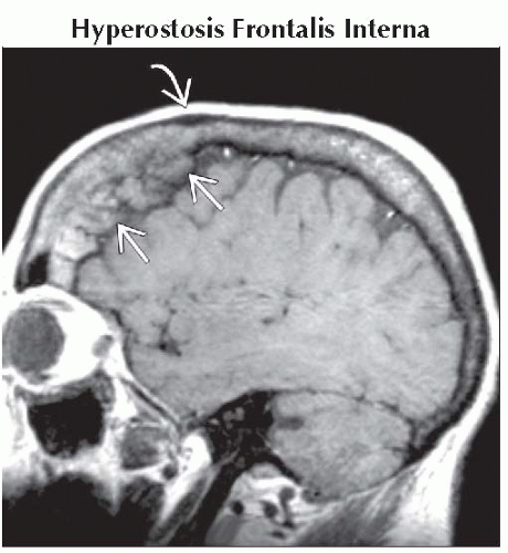 Sagittal T1WI MR shows a typical example of focal skull thickening from benign hyperostosis
 . Note that the thickening stops at coronal suture . Note that the thickening stops at coronal suture  . .Related posts:Stay updated, free articles. Join our Telegram channel
Full access? Get Clinical Tree
 Get Clinical Tree app for offline access

|
