(1)
Marina Spine Center, Marina del Rey, CA, USA
The 3rd rib resection is used for the transthoracic approach to the T1–T4 area. Resection of the 3rd rib allows greater spreading of the intercostal area than does 2nd rib resection [1]. The cephalad extension of the exposure is enhanced with kyphosis deformity of the cervicothoracic junction area. Exposure of the 3rd rib allows additional removal of the 2nd rib if the operative exposure is inadequate.
1.
Place the patient in the lateral decubitus position with the left side up. Prep and drape the entire left upper extremity, draping it sterilely out of the operative field.
2.
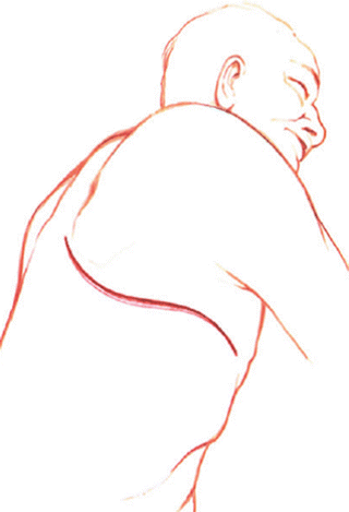
Incise the skin and subcutaneous tissue from the lateral paraspinous area at T1, along the medial caudal border of the scapula, under the axilla to the costal cartilage of the 3rd rib (Fig. 16.1).


Fig. 16.1
Place the patient in the lateral decubitus position with the left side up. Prep the entire extremity. Drape it out of the sterile operative field. Incise the skin and subcutaneous tissue from the lateral paraspinous area of T2 under the scapula to the costal margin of the 3rd rib
3.
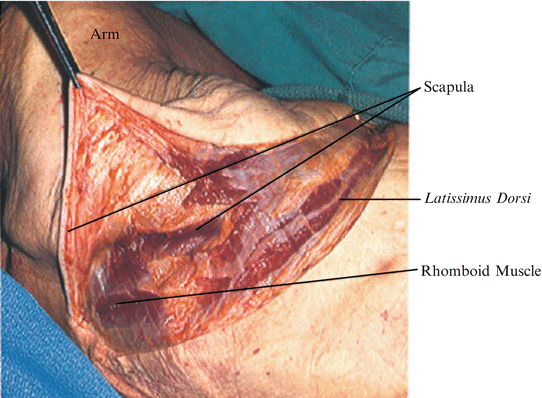
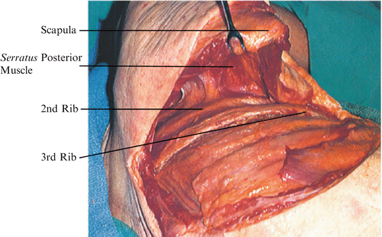
Carefully divide each subsequent muscle layer down to the level of the rib, sectioning portions of the trapezius, latissimus dorsi, rhomboid major, and serratus posterior (Fig. 16.2, 16.3), as needed. Careful dissection with electrocautery and meticulous cauterization of each muscle bleeding point allows exposure to the outer periosteum of the 3rd rib with a minimal amount of bleeding. As the muscle layers are divided, retract the scapula cephalad and medially to tense the muscle tissue for easier cutting. Palpate cephalad for identification of the 3rd rib (Fig. 16.3). Remember that the 1st rib is situated inside the 2nd; this is important for reaching the correct rib level.


Fig. 16.2
Carefully divide with electrocautery each subsequent muscle area down to the level of the 3rd rib. Section portions of the trapezius, latissimus dorsi, rhomboid major, and posterior serratus as needed. Control the intermuscular bleeding points

Fig. 16.3
Elevation of the scapula aids in the division of the muscles attached to the scapula and allows visualization of the 3rd rib. Palpate cephalad on the chest wall for positive identification of the 3rd rib. Remember, the 1st rib is located inside the 2nd rib and is sometimes missed
4.


For pathology such as large tumors of the cervicothoracic junction, several ribs, including the 1st can be resected, allowing for a large working area to C6. The limiting factor is that a stable rib must be left for support of the scapula [2].
Stay updated, free articles. Join our Telegram channel

Full access? Get Clinical Tree


