Third Ventricle Mass, Body/Posterior
Gregory L. Katzman, MD, MBA
DIFFERENTIAL DIAGNOSIS
Common
Pulsatile CSF
Dilated Suprapineal Recess
Neurocysticercosis
Less Common
Germinoma
Prominent Massa Intermedia, Chiari 2
Choroidal Metastases
Choroid Plexus Papilloma
Rare but Important
Xanthogranuloma
Ependymal Cyst
ESSENTIAL INFORMATION
Key Differential Diagnosis Issues
True primary posterior 3rd ventricle masses rare
Most represent extension from pineal pathology
Helpful Clues for Common Diagnoses
Pulsatile CSF
2° to time-of-flight effects/turbulent flow
↑ With thinner slices, longer TE, imaging perpendicular to flow
Evaluate other planes for real vs. artifact
Dilated Suprapineal Recess
Chronic aqueductal stenosis (any etiology)
Third ventricle dilates
May deform rostral tectum, mimic tectal glioma
Neurocysticercosis
Cystic lesion typically slightly hyperintense to CSF
± Discrete eccentric scolex
Cisterns > parenchyma > ventricles
Helpful Clues for Less Common Diagnoses
Germinoma
Usually extension from pineal tumor
Strong enhancement, ± CSF seeding
Restricted diffusion due to high cellularity
Prominent Massa Intermedia, Chiari 2
Large massa intermedia typical of Chiari 2
Choroidal Metastases
T1 hypo T2 hyperintense; avidly enhance
Lateral ventricles > 3rd > 4th
Choroid Plexus Papilloma
Strongly enhancing, lobulated mass
Hydrocephalus, ↑ intracranial pressure 2° to increased CSF production
Lateral ventricle > > 3rd
Helpful Clues for Rare Diagnoses
Xanthogranuloma
CT variable
MR T1 iso-hyper/T2 hyperintense
Lateral > > 3rd ventricle
Obstruction infrequent (3rd > lateral)
Ependymal Cyst
Nonenhancing thin-walled cyst
CSF density/intensity
Rare in 3rd ventricle
Image Gallery
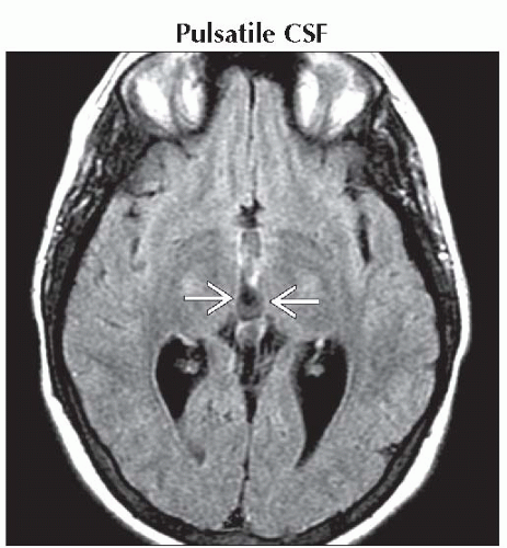 Axial FLAIR MR shows CSF flow anomaly
 manifesting as a hypointense “pseudolesion” of the posterior 3rd ventricle. Examining other sequences & planes confirmed this as flow artifact. manifesting as a hypointense “pseudolesion” of the posterior 3rd ventricle. Examining other sequences & planes confirmed this as flow artifact.Related posts:Stay updated, free articles. Join our Telegram channel
Full access? Get Clinical Tree
 Get Clinical Tree app for offline access

|
