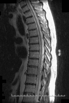
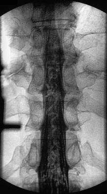
82 A 75-year-old man complained of increasing difficulty walking. He had a sensory level at T10-T11. T2-weighted thoracic magnetic resonance imaging (MRI) (Fig. 82-1) showed multiple flow voids in the dorsal aspect of the thoracic canal. A myelogram (Fig. 82-2) revealed a thoracic block with the presence of serpentine vessels. FIGURE 82-1 MRI with multiple flow voids seen in the dorsal aspect of the thoracic canal. A spinal angiogram (Fig. 82-3) showed a lower thoracic arteriovenous fistula with a feeding artery stemming from a right T7 radial artery. The artery of Adamkiewicz was identified on the left at T12. Type I spinal arteriovenous fistula
Thoracic Arteriovenous Fistula
Presentation
Radiologic Findings

Diagnosis

Thoracic Arteriovenous Fistula
Only gold members can continue reading. Log In or Register to continue

Full access? Get Clinical Tree


