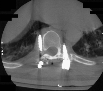
60 A 53-year-old man with known metastatic lung cancer to the spine complained of pleuritic chest pain following a transpedicular approach for a T7 corpectomy and T5-T9 pedicle screw fixation and arthrodesis. Computed tomography (CT) of the thoracic spine shows pedicle screw misplacement (Fig. 60-1). FIGURE 60-1 Axial CT of the thoracic spine shows misdirected thoracic pedicle screws. Extrapedicular screw placement The patient underwent revision surgery. When compared with a hook and rod construct, posterior thoracic fixation using pedicle screws provides instant three-column rigid fixation using a relatively short construct. This construct allows unloading of the incompetent ventral elements in patients too sick to undergo ventral or 360-degree surgery. Pedicle screws are also valuable in delayed kyphosis cases when the lamina has already been removed, obviating the use of hook or sub-laminar wires. Thoracic pedicle cannulation is contraindicated in pathologically compromised or anatomically unfit (“almond-shaped”) pedicles. Variability exists among spine surgeons as to the best trajectory for pedicle cannulation. Knowing the level, shape, and size of the pedicle, its coronal and sagittal relationship to the rest of the spine, as well as the surrounding anatomy is prerequisite for performing a safe pedicle screw placement. CT imaging and careful preoperative planning is essential. Intraoperative image guidance or fluoroscopy may further enhance pedicle screw placement. Belmont PJ Jr, Klemme WR, Dhawan A, Polly DW Jr. In vivo accuracy of thoracic pedicle screws. Spine 2001;26:2340–2346
Thoracic Screw Misplacement
Presentation
Radiologic Findings

Diagnosis
Treatment
Discussion
SUGGESTED READING
< div class='tao-gold-member'>
Thoracic Screw Misplacement
Only gold members can continue reading. Log In or Register to continue

Full access? Get Clinical Tree


