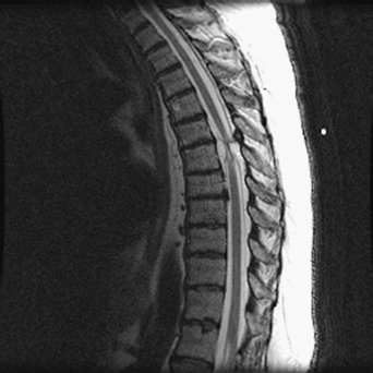
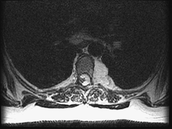
58 A 57-year-old woman had long-standing thoracic pain. She had brisk reflexes, but no other neurologic signs. An extensive medical workup was significant for a mass extending from the spinal canal, through the foramen, and under her sixth rib. T2-weighted magnetic resonance imaging (MRI) demonstrated a hyperintense soft tissue mass displacing the thoracic cord and extending through the neural foramen (Figs. 58-1 and 58-2). FIGURE 58-1 Sagittal MRI of the thoracic spine reveals a soft tissue mass displacing the cord. The pathologic specimen, a mixture of adipose and vessels, was an angiolipoma A T6 lateral extracavitary approach was performed for lesion excision. Although the typical spinal intraforaminal lesions are schwannomas, neurofibromas, and sometimes meningiomas, rarer lesions should be considered in the differential. An angiolipoma is a rare benign fatty tumor composed of mature lipocytes and multiple areas of angiomatous elements. It can be noninfiltrating, located in the subcutaneous tissue, or infiltrating, found in the deep soft tissue. It is benign and can be cured by surgical excision.
Thoracic Tumor
Presentation
Radiologic Findings
Diagnosis
Treatment
Discussion
Thoracic Tumor
Only gold members can continue reading. Log In or Register to continue


Full access? Get Clinical Tree


