TIAs and Strokes

Chapter 11 TIAs and Strokes


Transient ischemic attacks (TIAs) and strokes (or cerebrovascular accidents [CVAs]) cause readily recognizable constellations of transient or permanent neurologic deficits. Well-informed psychiatrists should be able to recognize their physical manifestations and anticipate their most common accompanying neuropsychologic manifestations, including amnesia, dementia, depression, and altered levels of consciousness. Psychiatrists should also be able to distinguish TIAs and strokes from conditions that produce similar clinical manifestations, such as seizures and brain tumors.


Transient Ischemic Attacks


As their name suggests, TIAs are temporary interruptions in cerebral circulation that give rise to neurologic deficits. Although the maximum allowable duration of a TIA is 24 hours, only 10% last longer than 4 hours and most resolve in 30–60 minutes. During that first hour of a neurologic deficit, neurologists who witness it cannot predict whether the deficit will completely resolve or become permanent. In other words, if the deficit resolves, it constitutes a TIA, but if the deficit persists, it constitutes a stroke. Most of the time neurologists diagnose TIAs only in retrospect.


The majority of TIAs result from platelet emboli that have formed on the surface of atherosclerotic plaques that have built up on the inner wall of the extracranial arteries: the carotid and vertebral arteries and the aortic arch. The plaques are usually ulcerated and cause some degree of stenosis. When platelet emboli leave their surface, they course through a cerebral artery, temporarily interrupt a portion of the cerebral circulation, and induce a brief period of ischemia. Alternatively, cardiac arrhythmias and other causes of hypotension produce TIAs.


Not only do TIAs cause temporary neurologic deficits, they also reflect underlying atherosclerotic cerebrovascular disease and represent a risk factor for stroke. TIAs lead to strokes when either an atherosclerotic plaque grows large enough to occlude an extracerebral vessel or it throws off an embolus that permanently blocks a “downstream” cerebral artery. Within the first year following a TIA, approximately 12% of patients develop a stroke. Each year thereafter, an additional 5% develop one.


TIAs mimic other transient neurologic conditions, particularly partial seizures, postictal confusion and (Todd’s) hemiparesis, hemiplegic migraine, metabolic aberrations, adverse reactions to medicines, and, rarely, effects of cerebral mass lesions. In addition, when TIAs produce aphasia, amnesia, or another neuropsychologic deficit, but no physical deficit, they mimic psychogenic episodes.


Carotid Artery TIAs


Platelet emboli that form on plaques at the common carotid artery bifurcation (Fig. 11-1) can lead to cerebral-hemisphere TIAs. Contralateral hemiparesis, hemisensory loss, paresthesias, or homonymous hemianopsia characterize carotid artery TIAs. Depending on whether the dominant or nondominant carotid artery gives rise to the problem, a TIA may induce neuropsychologic aberrations accompanied or unaccompanied by hemiparesis and other physical deficits. For example, dominant-hemisphere TIAs may suddenly, but briefly, cause aphasia with or without hemiparesis or homonymous hemianopsia. Similarly, nondominant-hemisphere TIAs may cause brief neglect or hemi-inattention with or without homonymous hemiparesis.


image

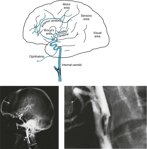
FIGURE 11-1 Top, At its bifurcation in the neck, the common carotid artery divides to form the external and internal carotid arteries. Giving off no branches until it is within the skull, the internal carotid artery immediately sends off the ophthalmic artery. The internal carotid artery then divides into the anterior and middle cerebral arteries. It also gives rise to the posterior communicating artery – not the posterior cerebral artery. Thus, each internal carotid artery perfuses the ipsilateral eye and anterior and middle portions of the ipsilateral cerebral hemisphere. The middle cerebral artery, a major branch of the internal carotid artery, supplies the deep and mid-section of the hemisphere. This region contains most of the motor cortex, sensory cortex, and, in the dominant hemisphere, the perisylvian language arc (see Fig. 8-1). Each anterior cerebral artery, another branch of the internal carotid artery, supplies the frontal lobe, including the medial surface of the motor cortex, which contains the motor innervation for the leg. The posterior cerebral artery (see Fig. 11-2), terminal branches of the basilar artery, supplies the occipital lobe, which houses the visual cortex, and most of the temporal lobe. Bottom left, An arteriogram of the carotid artery and its branches prominently displays the bifurcation (black arrow), the typical “candelabra” of branches that comprise the middle cerebral artery, and a faint anterior cerebral artery that sweeps from anterior to posterior (white arrow). Bottom right, A magnification of the bifurcation reveals an extensive circumferential plaque constricting the internal carotid artery. The remaining blood flow appears as an “apple core.” The rough interior surface of the artery gives rise to retinal and cerebral emboli.


Sometimes a TIA causes only brief visual loss in one eye, i.e., monocular blindness. Neurologists call this distinctive symptom amaurosis fugax (Greek, fleeting darkness). The underlying mechanism consists of emboli from the internal carotid artery flying into the ophthalmic artery, the carotid artery’s first branch, to induce several minutes of ischemia in the retina and optic nerve (Box 11-1). Unlike migraines, which can also cause transient visual loss, TIAs rarely cause headache or scintillations.


Between each TIA, patients have no lasting neurologic deficits. However, occasionally auscultation over the carotid artery bifurcation reveals a harsh systolic sound (bruit) that suggests carotid artery stenosis or at least atherosclerotic cerebrovascular disease. Retinal emboli (Hollenhorst plaques) of atheromatous material, which neurologists see on fundoscopy, strongly suggest carotid artery atherosclerotic plaque with stenosis.


In patients with mild cognitive impairment, TIAs may produce a brief but marked confusional state. A similar disturbance may occur after atherosclerosis slowly occludes one carotid artery, forcing the other carotid artery to supply both cerebral hemispheres through the circle of Willis (Fig. 11-2, top right). In this case, the cerebral blood supply is tenuous and emboli from the patent artery produce bilateral cerebral ischemia.


image

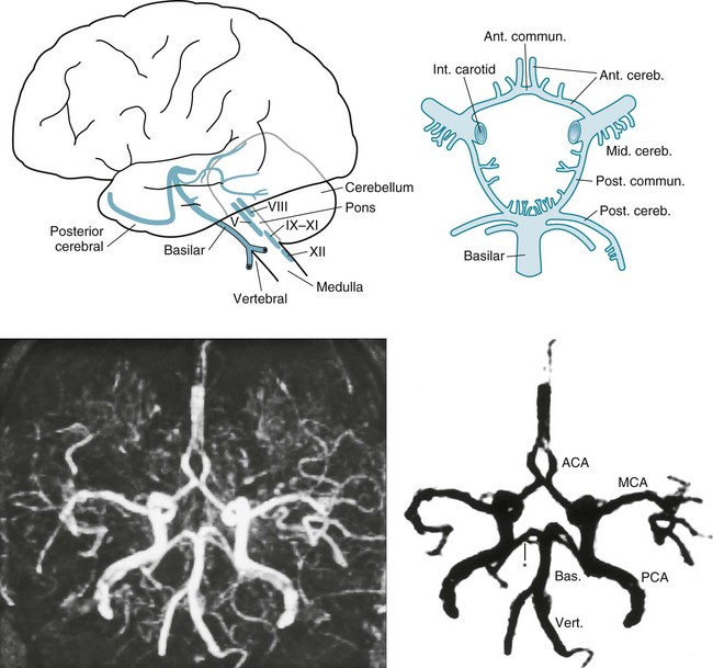
FIGURE 11-2 Top left, After ascending, encased in the cervical vertebrae, the two vertebral arteries enter the skull. They join to form the basilar artery at the base of the brain. Small, delicate branches of the basilar artery supply the brainstem and its contents. (The Roman numerals refer to cranial nerve nuclei.) Large branches, as if wrapping their arms around the brainstem, supply the cerebellum and posterior portion of the cerebrum (i.e., the occipital lobes and inferomedial portions of the temporal lobes). The posterior cerebral arteries are, for practical purposes, the terminal branches of the basilar artery. They supply the occipital cortex and the posterior, inferior aspect of the temporal lobes. Top right, Belying its name, the circle of Willis, the “great anastomoses,” is completely patent in only about 20% of people. Connections between the basilar and internal carotid arteries ideally form the circle, which should give off the anterior, middle, and posterior cerebral arteries. The circle should provide anastomoses between anterior–posterior and right–left cerebral circulations. Despite the advantages that the circle confers, junctions of the arteries are weak spots. Defects at the junctions may balloon outward to form berry aneurysms that rupture and produce subarachnoid hemorrhages. Bottom left, This axial magnetic resonance angiogram (MRA) shows the major cerebral arteries that form the circle of Willis and anterior and posterior circulations. Bottom right, This MRA highlights the major cerebral arteries and shows the anterior and posterior circulations. The anterior circulation consists of the middle cerebral arteries (MCA), anterior cerebral arteries (ACA), and the anterior communicating artery (∧), which joins the ACAs. The posterior circulation consists of the basilar (Bas.) artery and two vertebral (Vert.), posterior cerebral (PCA), and posterior communicating arteries (*), which connect the PCAs and the basilar artery.


Preventive Measures


The goal of treatment is not merely to prevent recurrences of TIAs, but to prevent a stroke. In conjunction with the patient’s internist, neurologists generally suggest stroke risk reduction measures, such as smoking cessation, weight reduction, and treatment of hypertension, diabetes, and elevated cholesterol. In addition, neurologists usually suggest inhibiting platelet aggregation with aspirin (81 mg daily), clopidogrel (Plavix), or a dipyridamole–aspirin combination (Aggrenox). They usually suggest warfarin (Coumadin) or other anticoagulants to prevent cardioembolic embolization in patients with atrial fibrillation, left ventricular thrombus, and certain other cardiac conditions.


For TIA patients with atherosclerotic plaque causing at least 70% stenosis of the common or internal carotid artery, neurologists often recommend a carotid endarterectomy. This is invasive, delicate surgery in which surgeons briefly open the artery to remove the plaque. Although effective, carotid endarterectomy carries substantial risk because it briefly interrupts the cerebral blood supply and potentially allows pieces of the plaque to enter the cerebral circulation that, like emboli, may cause a stroke. Carotid endarterectomy for asymptomatic individuals with comparably severe carotid stenosis may also be indicated to prevent a stroke; however, the criteria remain uncertain. No procedure is feasible for a completely occluded artery.


For patients who cannot undergo carotid endarterectomy, an alternative is intravascular insertion of stents. These devices are essentially expandable tubes that neuroradiologists insert intravascularly into the carotid artery to widen atherosclerotic stenoses (see Fig. 20-28). They also trap underlying atheromatous debris against the inner surface of the arterial wall to reduce the likelihood of emboli. In contrast to the benefits of inserting stents into the extracranial carotid arteries, inserting stents into intracerebral arteries, such as the basilar or middle cerebral artery, carries too much risk to justify its use.


Basilar Artery TIAs


The vertebrobasilar system, basilar artery system, or simply the posterior circulation supplies the brainstem, cerebellum, and the posterior inferior portion of the cerebrum (the occipital and medial inferior portion of the temporal lobes [see Fig. 11-2]). Emboli-generating plaques tend to develop at both the origin of the vertebral arteries (in the chest) and their junction at the base of the brain.


Symptoms and signs of basilar artery TIAs, which usually result from patchy brainstem ischemia, differ greatly from those of carotid artery TIAs (Box 11-2). Typical basilar artery TIA symptoms include tingling around the mouth (circumoral paresthesias), dysarthria, nystagmus, diplopia, ataxia, and vertigo. On rare occasions, when all blood flow through the basilar artery momentarily stops, almost the entire brainstem suffers from ischemia. The brainstem ischemia interrupts consciousness and body tone, which causes patients to collapse. This TIA, termed a drop attack, strikes suddenly and unexpectedly. (It appears similar to cataplexy [see Chapter 17].)


Vertigo represents one of the most characteristic – but perplexing – symptoms of basilar artery TIAs. As a medical symptom, vertigo means a sensation of the patient or the surroundings revolving or otherwise moving. The thoughtful physician should accept no other descriptions. In particular, the common complaint of “dizziness” has no clinical value because, depending on the patient, it may mean imbalance, lightheadedness, anxiety, confusion, or impending trouble.


The evaluation of basilar artery symptoms typically includes – as with the evaluation of carotid artery symptoms – MRI, MRA, and evaluation for cardiac and systemic illness. In addition, a transcranial Doppler examination, which harmlessly penetrates the skull, may portray the vertebrobasilar system’s architecture. Neurologists rely on the same medications used for carotid artery TIAs. Because the usual sites of vertebrobasilar stenosis remain shielded by the chest, vertebrae, and skull, an endarterectomy would not be feasible.


Transient Global Amnesia


TIAs sometimes impair the circulation of the basilar artery’s terminal branches, the posterior cerebral arteries, which supply the temporal lobes (see Fig. 11-2). Because the temporal lobes contain portions of the limbic system, particularly the hippocampi (see Fig. 16-5), posterior circulation TIAs may induce episodes of temporary amnesia and personality change – called transient global amnesia (TGA).


The fundamental feature of TGA is an acutely developing period of amnesia. During an attack, patients cannot memorize or learn new information, such as a sequence of digits, i.e., they have anterograde amnesia. They also cannot recall recently acquired information, such as the events of the last several hours or days, i.e., they have retrograde amnesia. Although patients suffer both antero- and retrograde amnesia, the anterograde amnesia is more profound, has a greater duration, causes the greater impediment, and creates more distress. As examples of their anterograde amnesia, patients typically do not know how they came to the physician’s office or the emergency room. Once there, they lose track of their responses during their examination by a physician, who may require reintroduction several times during the examination.


In the midst of TGA, individuals may perform rote but relatively complex activities, such as driving a well-known route or preparing dinner for a large family. However, unable to comprehend their situation, some individuals become distraught, agitated, or panicked. Some, as if recoiling, appear apathetic and immobile. Most, however, seem calm but bewildered.


TGAs typically occur in middle-aged and older individuals who are apt to have cerebrovascular disease. These attacks typically develop in the midst of frightening events or physical exertion, particularly sexual activity – a coincidence that might lead to misinterpretation.


In contrast to their amnesia for new facts, TGA patients characteristically retain their general knowledge and fundamental personal information. For example, they remain able to recite their name, address, telephone number, and occupation. (All this preserved memory contradicts the adjective global in TGA.)


After their abrupt onset, TGAs last for 3–24 hours, but their intensity is most pronounced during the initial 1–2 hours. By definition, the total duration must not exceed 24 hours. The recurrence rate is approximately 10% and they do not represent a risk factor for stroke. Neurologists have proposed various etiologies for TGA, including migraine, complex partial seizures, metabolic aberrations, use of certain medicines, and congestion of cerebral veins, as well as posterior-circulation TIAs. Although the posterior-circulation TIA theory remains the most popular, TGAs do not behave like TIAs. For instance, TGA rarely if ever recurs, develops in conjunction with TIAs in other arterial distributions, shows TIA-like MRI changes, or precedes a stroke.


Even in the absence of a confirmatory laboratory test for TGA, its clinical features differentiate it from other neurologic conditions. TGA patients’ preserved intellect and general knowledge, as well as their remaining fully conscious, distinguishes them from patients in delirium. TGA patients, despite their amnesia, also do not confabulate in the manner of Wernicke–Korsakoff patients. Complex partial seizures differ by producing dulling of the sensorium, simple repetitive actions, paroxysmal or other epileptiform EEG changes, and a high rate of recurrence (see Chapter 10).


Physicians using the preliminary version of the Diagnostic and Statistical Manual of Mental Disorders, 5th edition (DSM-5) might be tempted to diagnose a patient with no recall of recent events as having Dissociative Amnesia, which it defines primarily as “inability to recall important autobiographical information, usually of a traumatic or stressful nature” and requires that, presumably over time, patients experience “significant distress or impairment in social, occupational, or other important areas of functioning.” However, that diagnosis would exclude TGA patients because they retain fundamental personal information. The preliminary version of DSM-5 also notes a subtype of Dissociative Amnesia, Dissociative Fugue, previously called Psychogenic Fugue, in which individuals purposefully travel or show “bewildered wandering.” Presumably, after establishing a new location, these individuals function within it and do not recall their previous life’s crucial aspects, such as a spouse, debts, or crimes. In other words, unlike TGA patients, these patients’ anterograde memory remains normal, but their retrograde amnesia persists, and the disorder lasts for months, years, or possibly indefinitely.


Strokes


Strokes cause permanent physical and neuropsychologic deficits. Most result from an arterial thrombosis, embolus, or hemorrhage. Feeling that there is nothing accidental about strokes, neurologists have largely abandoned the term “cerebrovascular accident.”


Risk Factors


Reflecting the necessity of preventing strokes, epidemiologic studies constantly search for stroke risk factors (Box 11-3). Although physicians customarily list stroke risk factors as individual threats, the factors actually tend to cluster. For example, obesity, elevated low-density lipoprotein cholesterol, and hypertension frequently occur together in the metabolic syndrome. Some factors, by themselves, have a weak correlation with stroke, but pose a synergistic risk when paired with others. For example, cigarette smoking correlates with stroke; however, with concomitant use of oral contraceptives or in sufferers of migraine with aura, it becomes a moderately powerful risk factor. More strikingly, smokers who are hypertensive have a 20-fold greater risk. Curiously, total serum cholesterol carries a strong risk for myocardial infarctions but a weak risk for stroke.


Age older than 65 years constitutes a powerful risk factor. Yet, about 25% of stroke victims are younger than 65 years and 12% younger than 45 years. Stroke even occurs in adolescents and young children. Hypertension, another major risk factor, similarly leads to strokes in younger individuals as well as in adults. It is also probably the most common cause of stroke-induced dementia.


Various cardiac conditions – valvular disease, prosthetic valves, acute myocardial infarction, and atrial fibrillation – comprise another risk factor because they tend to produce thromboses on valves and endocardial surfaces that embolize to the brain and elsewhere. Treatment with an anticoagulant greatly reduces the incidence of embolic stroke in patients with these cardiac diseases.


Diabetes mellitus and elevated total cholesterol represent powerful risk factors for myocardial infarction but less so for stroke. In fact, for individuals younger than 45 years, elevated total cholesterol carries little risk of stroke.


Cigarette smoking and inhaling second-hand smoke convey a significant risk factor for stroke. Even former cigarette smokers retain an almost twofold greater risk of stroke. Although judicious alcohol drinking (one drink daily to weekly) provides a slight protective effect, heavy alcohol intake poses a risk.


Migraine – in general – represents a risk factor, but one with minor significance. Any increased risk is probably restricted to migraineurs who smoke, use oral contraceptives, or have migraine with aura. Drug abuse frequently causes strokes through intravenous injection of particulate material, episodes of anoxia and hypotension, cerebral vasculitis, hypertension, and vasospasm. For example, amphetamines and cocaine are sympathomimetic stimulants that induce bursts of hypertension and prolonged arterial vasospasm that routinely lead to stroke or myocardial infarction. In particular, cocaine alkaloid (“crack cocaine”) notoriously leads to cerebral hemorrhage. By way of contrast, marijuana intoxication or even its chronic use is not associated with strokes. Studies have also implicated over-the-counter medicines, particularly phenylpropanolamine and ephedra, which are sympathomimetic ingredients of weight loss and cough suppressant medicines. Because of their association with strokes, the Food and Drug Administration has banned many of these medications.


Estrogen, when used alone as for postmenopausal hormone replacement, slightly increases the stroke risk. The danger from oral contraceptives is probably restricted to the original, high-dose estrogen preparations. Currently available low-dose estrogen preparations confer only a negligible risk for stroke.


Elevated serum concentration of homocysteine serves as a marker for an increased risk. In the autosomal recessive genetic condition, homocystinuria, children have a Marfan-like habitus, ocular lens displacement, and other anatomic abnormalities. More importantly, they routinely suffer strokes in childhood. Elevated homocysteine levels in adults are associated with the use of antiepileptic drugs and coronary artery and peripheral vascular disease, as well as stroke. Folic acid reduces elevated serum homocysteine concentrations (see Fig. 5-8), as well as reducing the incidence of fetal neural tube defects (see Chapter 13), but surprisingly, it does not reduce the incidence of strokes.


Although most risk factors relate to atherosclerosis, credible data suggest that systemic infection or vascular inflammation can also give rise to strokes. For example, elevated serum concentrations of a marker for inflammation, C-reactive protein, has a strong association with strokes. Other compelling data link periodontal disease to subsequent stroke.


The presence of anticardiolipin antibodies constitutes another example of systemic inflammation constituting a risk factor for stroke. These antibodies are a hallmark of the antiphospholipid syndrome. This syndrome is a hypercoagulable state in which mostly young women develop repeated deep vein thromboses (DVT), suffer miscarriages, and have a susceptibility to migraine and stroke. Treatment, mostly aimed at preventing thromboses, consists of anticoagulants and sometimes immunosuppressives. Although the antiphospholipid syndrome carries no specific direct psychiatric comorbidity, its associated migraines and repeated miscarriages have psychiatric repercussions. Thus, a young woman who seeks psychiatric consultation for depression following two or more miscarriages and has a history of migraine and DVT may actually have the antiphospholipid syndrome.


Examples abound of hematologic disorders causing strokes. For example, sickle cell disease and, under certain circumstances, sickle cell trait lead to strokes. Because pregnancy also induces a hypercoagulable state, it causes a small but significantly increased incidence of strokes during pregnancy, delivery, and the postpartum period. In addition, several obstetric problems, such as eclampsia, venous sinus thrombosis, cortical vein thrombosis, and ruptured aneurysms, lead to stroke-like brain damage.


Surgery is a risk factor. Not surprisingly, cardiac surgery is riskier than noncardiac surgery, urgent surgery is riskier than elective surgery, and older surgery patients are more vulnerable than younger ones.


Thrombosis and Embolus


Thromboses may simply occlude cerebral arteries and deprive the “downstream” brain of its blood flow. Similarly, emboli from the heart, aortic arch, cervical extracranial arteries, or intracranial arteries may lodge in a cerebral artery and disrupt cerebral blood flow. Because cerebral thromboses and emboli deprive a region of the brain of its oxygen supply, they cause ischemic infarction. Comprising up to 85% of all strokes, cerebral ischemic infarctions are by far the most common form.


Other important conditions that lead to stroke include vasculitis, drug abuse, sickle cell disease, and other blood dyscrasias. In short, the most frequent cause of stroke consists of an abnormality of the heart, blood vessels, or blood.


As if Napoleon’s dictum “Geography is destiny” lives, present-day neurologists readily explain that the interrupted vascular supply lines of a stroke determine neurologic deficits. The time course of a stroke suggests whether it originated in an embolus or a thrombosis. Cerebral emboli-induced infarctions develop suddenly as the embolus lodges in a cerebral vessel. Usually deficits are maximal at the onset of an embolic stroke and resolve to a certain extent over the next several days. Cerebral thromboses, in contrast, generally develop slowly or intermittently and often begin during sleep. With both embolic and thrombotic strokes, the region surrounding the infarction becomes edematous. In large strokes, edema is most severe and neurologic deficits are most pronounced during the third to fifth days. Some clinical improvement occurs as the edema resolves and ischemic areas recover; however, the infarction remains a functionless scar.


In addition to creating permanent clinical deficits, stroke scars are potentially epileptogenic. Approximately 50% of seizures that develop in adults older than 65 years originate from these lesions (see Chapter 10). Thus, patients with a history of having sustained a stroke who develop confusion, unresponsiveness, or abnormal behavior may have suffered a complex partial seizure as well as another stroke or TIA.


Necrosis


Ischemic strokes, like brain tumors, gunshot wounds, and other destructive processes, lead to cell death by necrosis. Cellular necrosis requires no cellular energy to begin or progress, but the necrotic debris (dead cells) elicits a cellular inflammatory response. In contrast, cell death by apoptosis takes place in amyotrophic lateral sclerosis (ALS), Huntington disease, and many other neurodegenerative diseases. Apoptosis also serves as the mechanism for the normal involution of several organs, including the thymus gland and ductus arteriosus. Unlike necrosis, apoptosis requires cellular energy to begin and progress, and the dead cells do not evoke an inflammatory response.


Infarctions in the Carotid Artery Distribution


Cerebral artery thrombosis and embolism produce an infarction in the distribution of the occluded artery. These strokes result in well-known clinical deficits (see Fig. 11-1 and Box 11-4):


Infarctions in the Basilar Artery Distribution


Infarctions in the basilar artery distribution cause brainstem, cerebellar, or posterior cerebral injuries. Small brainstem infarctions usually cause constellations of cranial nerve injuries and hemiparesis, and large ones usually cause coma, if not immediate death. In contrast to cerebral-hemisphere infarctions, brainstem infarctions generally spare language or intellectual function and do not cause seizures (see Box 11-4).


The posterior cerebral arteries, as previously noted, are actually terminal branches of the basilar artery. Infarction of a posterior cerebral artery causes a contralateral homonymous hemianopsia and occasionally alexia without agraphia (see Chapter 8). Bilateral posterior cerebral artery strokes can cause (cortical) blindness, which sometimes leads to Anton syndrome (see Chapter 12).


Neurologists love to localize small brainstem infarctions for both clinical and academic reasons. Lesions of the midbrain cause ipsilateral oculomotor nerve and contralateral paresis (see Fig. 4-9). Those in the pons cause ipsilateral abducens nerve and contralateral paresis (see Fig. 4-11). Infarctions involving the midline of the midbrain or pons cause the medial longitudinal fasciculus syndrome (see Chapters 12 and 15). Finally, lateral medullary infarctions, which are the most common brainstem infarction, cause a complex, apparently disparate, combination of ipsilateral limb ataxia, palatal paresis, Horner syndrome, and alternating hypalgesia (see Wallenberg syndrome, Fig. 2-10).


The clinical implication of such precise localization indicates that, if the lesion is situated in the brainstem, the patient’s cognitive functions will remain intact. For example, a patient with right hemiparesis and a left sixth cranial nerve palsy is unlikely to have aphasia or dementia. Even extensive, devastating brainstem lesions will spare intellectual function and preserve cognition – as in the locked-in syndrome (see later).


Hemorrhages


Cerebral or cerebellar hemorrhages typically occur abruptly and, because of increased intracranial pressure, produce the triad of headache, nausea, and vomiting. Patients usually lose consciousness and have profound neurologic deficits determined by the hemorrhage’s location. One special example is the cerebellar hemorrhage because it potentially leads to rapidly fatal compression of the fourth ventricle and the underlying medulla. Compression of the fourth ventricle blocks the flow of cerebrospinal fluid (CSF), which causes obstructive hydrocephalus. Pressure on the medulla depresses respiratory drive and causes coma. Physicians can diagnose cerebellar hemorrhage by its clinical manifestations – occipital headache, gait ataxia, dysarthria, and lethargy. Once imaging studies confirm a diagnosis of cerebellar hemorrhage with hydrocephalus, neurosurgeons often immediately evacuate the hemorrhage, insert a ventricular shunt, or both to relieve the pressure on the fourth ventricle and brainstem.


Hemorrhages, which are most often the result of hypertension, usually erupt in the basal ganglia, thalamus, pons, or cerebellum (see Figs 20-13 and 20-14). Trauma and use of cocaine also cause hemorrhages, but their location is not as predictable as with hypertension-induced ones. In a classic problem, patients who take a monamine oxidase inhibitor (MAOI) antidepressant and then, inadvertently or in a suicide attempt, consume certain foodstuffs, notoriously aged cheese or red wine, or receive meperidine (Demerol), develop cerebral hemorrhages. These combinations, which are sympathomimetic, lead to a burst of hypertension sufficient to cause the hemorrhage. (Notably, MAOI-A antidepressants may cause this problem, but MAOI-B medications, such as those used to treat Parkinson disease, generally do not [see Chapter 9].)


Subarachnoid hemorrhage (SAH), although usually traumatic in origin, sometimes results from a ruptured berry aneurysm. SAH most often produces a prostrating headache that patients classically describe as the “worst headache” of their life. SAH also produces nuchal rigidity and lethargy, but usually no lateralized signs. Exercising, straining at stool, and other usually benign activities can rupture an aneurysm and precipitate an SAH. A diagnostic dilemma occurs when a sudden, profound headache interrupts sex. In this case, the exertion may have led to an SAH, but more often coital cephalalgia is responsible (see Chapter 9). Thus, TGA, SAH, and a migraine variant are hazards of sexual activity.


When an aneurysm ruptures, CT and MRI usually reveal blood in the subarachnoid space at the base of the brain or within the ventricles. A lumbar puncture (LP) yields bloody or xanthochromic (yellow) CSF. Depending on the particular case, neurologists may order CT angiography, MRA, or conventional angiography to detect an aneurysm.


The traditional treatment of a ruptured aneurysm consisted of a craniotomy to clamp the neck of the aneurysm. Even for aneurysms located in accessible positions, surgical complications included vascular spasm, rupture of the aneurysm that had only been leaking, and inadvertent occlusion of the parent vessel. Current treatment options, which represent a major medical advance, include minimally invasive procedures, such as intravascular insertion of coils, epoxies, and other substances that fill the aneurysm. However, surgery remains the only option for aneurysms inaccessible by an intravascular approach.


Neuropsychologic Sequalae


Several large or many small (lacunar) strokes cause vascular cognitive impairment, previously labeled vascular dementia, and before that multi-infarct dementia. The preliminary version of the DSM-5 labels it Vascular Neurocognitive Disorder. Ironically, despite the implication of these terms, the dementia stems from the neuropathologic changes of Alzheimer disease that accompany those of cerebral vascular disease (see Chapter 7).


Occlusion of a cerebral artery typically destroys a discrete critical area of the cerebral cortex. Commonly occurring strokes produce well-known neuropsychologic syndromes, such as aphasia, Gerstmann syndrome, and hemi-inattention (see Chapter 8). Strokes of both frontal lobes, which may occur either simultaneously or in succession, cause pseudobulbar palsy and frontal lobe dysfunction (see Chapters 4 and 8).


In another mechanism, hypoxia, hypotension, carbon monoxide poisoning, or similar catastrophes cause generalized cerebral anoxia. This insult leads to dementia, cortical blindness, a persistent vegetative state (PVS: see later), and postanoxic myoclonus (see Chapter 18). A variation of generalized anoxia occurs when cerebral anoxia affects only the fine, terminal branches of cerebral arteries that perfuse the extensive circumferential regions of the cerebral cortex, which neurologists call the watershed or borderzone area. An episode of cerebral anoxia that reduces the oxygen supply below a critical point in the watershed area produces a watershed infarction

Only gold members can continue reading. Log In or Register to continue

Stay updated, free articles. Join our Telegram channel

Jun 4, 2016 | Posted by in NEUROLOGY | Comments Off on TIAs and Strokes

Full access? Get Clinical Tree

Get Clinical Tree app for offline access