Chapter 13 Introduction to low-frequency currents
INTRODUCTION
The term ‘low-frequency currents’ covers a range of electrical currents commonly used in clinical practice to stimulate muscles, nerves or a combination of both. The physiological effects of such stimulation are used therapeutically to strengthen muscle (Healy et al 2006, Stevens et al 2004), assist in wound healing (Bogie et al 2000, Kloth 2005), relieve pain (Khadilkar et al 2005) and reduce oedema (Man et al 2003). As the number of devices producing electrical currents for therapeutic applications has proliferated over the past few decades, the terminology relating to the output of these devices has become somewhat confusing. This introductory chapter will therefore provide an overview of the basic principles associated with these type of currents using standardized terminology; this will serve as a foundation for the subsequent chapters in this section that focus on individual currents: transcutaneous electrical nerve stimulation (TENS) and neuromuscular electrical stimulation. The chapter commences with a brief description of how an electrical current initiates an action potential in a nerve. This is followed by an explanation of the characteristics or stimulation parameters of the currents. Finally, a guide for the safe application of electrical currents is provided.
STIMULATION OF A NERVE USING ELECTRICAL CURRENTS
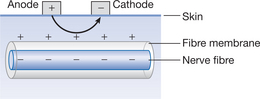
Chapter 5 describes the generation of an action potential in a nerve. This chapter gives a brief summary of stimulation of nerves using electrical currents as an introduction to the following chapters. An externally applied stimulus can cause depolarization of a nerve and thus initiate an action potential as long as the applied stimulus depolarizes the resting membrane potential to the threshold level. Depolarization means loss of the normal negative value of the resting membrane potential in a nerve (-270mV). In an electrical circuit, one electrode is positively charged (anode) and the other is negatively charged (cathode). In the circuit, the current flows from anode to cathode (Fig. 13.1). Under the cathode, negative charges accumulate on the outer surface of the nerve fibre membrane as they are repelled by its negative charge; the outside of the membrane becomes relatively more negative. The inside of the membrane therefore becomes more positive, causing depolarization. For this reason, the cathode is commonly termed the ‘activeelectrode’. Once the critical level of depolarization occurs (i.e. threshold level is reached), an action potential is initiated. Under the anode, negative charges move from the outside of the membrane towards the anode as they are attracted towards its positive charge. This makes the outside of the membrane more positive and therefore the inside of the membrane more negative, hence hyperpolarization occurs.
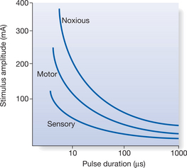
If the applied stimulus is electrical, the stimulation parameters of frequency, pulse duration and amplitude must correlate with the characteristics of the nerve fibre itself. The relationship between the stimulation parameters of pulse amplitude and duration of the stimulating current can be illustrated on a strength–duration (SD) curve. Basically, this curve illustrates the relationship between different combinations of pulse duration and amplitude that are required to optimally stimulate a specific fibre type and produce the response associated with the fibre, i.e. motor, sensory or noxious response. Figure 13.2 illustrates SD curves for motor, sensory and nociceptive fibres. At any given pulse duration, the order of recruitment with increasing stimulus amplitude is sensory, then motor, then nociceptive fibres.
STIMULATION PARAMETERS OF LOW-FREQUENCY CURRENTS
DOCUMENTATION OF PARAMETERS
One of the key factors associated with a poor treatment outcome is the use of inappropriate treatment parameters (Sluka & Walsh 2003). Inadequate documentation of the stimulation parameters in clinical notes or research papers makes it inherently difficult to replicate that study by other clinical colleagues/researchers. Several Cochrane Systematic Reviews have been published on the efficacy of low-frequency currents for a variety of conditions (Table 13.1). A common observation by the authors of these reviews is the lack of standardized treatment regimens, which makes comparisons across clinical trials an impossible task.
Table 13.1 Cochrane Systematic Reviews of low-frequency electrical currents
| Author and year | Title |
|---|---|
| Khadilkar et al 2005 | TENS for chronic low back pain |
| Cameron et al 2003 | TENS for dementia |
| Brosseau et al 2003 | TENS for treatment of rheumatoid arthritis of the hand |
| Proctor et al 2002 | TENS and acupuncture for primary dysmenorrhoea |
| Pelland et al 2002 | Electrical stimulation for the treatment of rheumatoid arthritis |
| Osiri et al 2000 | TENS for knee osteoarthritis |
| Hosker et al 2007 | Electrical stimulation for faecal incontinence in adults |
| Carroll et al 2000 | TENS for chronic pain |
The type of information required for documentation of any low-frequency current includes: type of current, frequency, pulse duration, amplitude, waveform, type of output, duty cycle, duration of treatment and electrodes (placement, size, shape). This section will briefly outline the basic stimulation parameters relevant to the electrical currents described in Chapters 14–18.


Stay updated, free articles. Join our Telegram channel

Full access? Get Clinical Tree


