(1)
Showa University School of Medicine, Tokyo, Japan
The trans-11th rib extrapleural approach is less traumatic to the respiratory system1 and has the advantage that a patient does not necessarily need either thoracic drainage or a postoperative intensive care unit (ICU) stay. The incidence of postoperative atelectasis is very rare, because ventilation is maintained and no direct mechanical pressure is applied to the lung tissue during the spinal procedure; this is beneficial for geriatric or ventilatory poor-risk patients. Even if the parietal pleura is accidentally perforated, suturing the injured pleura requires less intensive postoperative management compared to the ordinary thoracotomy procedure.
1.
The exposable cephalad operative field for 11th rib resection is the T11 vertebral body and the intervertebral disc of T11–T12. Likewise, caudad exposure is the L2 vertebral body. Pathology that has occurred around T12 or L1 is the most convenient target. If the skin incision is extended toward the symphysis pubica caudally, the L5–S1 disc space can be reached with this approach.
2.
Indications for this approach are determined by pathology that exists anterolateral to the following spinal levels:
Disc herniation at T11–T12, T12–L1, L1–L2
Burst fracture, tumor, spondylitis, or ossification of the posterior longitudinal ligament (OPLL) at T12, L1, L2
Scoliosis or kyphotic deformity at the thoracolumbar junction
3.
Contraindications for this approach include previous thoracotomy. Spinal infection, either pyogenic or tuberculous, or metastatic spinal tumor is not necessarily a contraindication unless severe adhesive pleuritis is expected. Rather, these diseases can be good indications because the parietal pleura plays a role in barricading disease dissemination.
The patient is placed in either right or left lateral decubitus position depending on convenience in approaching the spinal pathology. As in ordinary thoracotomy, the upper extremity of the open side is lifted forward and an axillar pad is placed under the dependent axilla. The operating table should be slightly bent to maintain the coronal spinal physiologic alignment.
Get Clinical Tree app for offline access

1.
In the same fashion as in ordinary trans-11th rib thoracotomy, a curved oblique incision is made over the 11th rib. The posterior border of this incision is placed just lateral to the margin of the paravertebral muscles, and the anterior border is 3 to 5 cm beyond the tip of the 11th rib (Fig. 21.1). The incision can be extended toward the symphysis pubica depending upon how far down the exposure to the lumbar spine is needed.
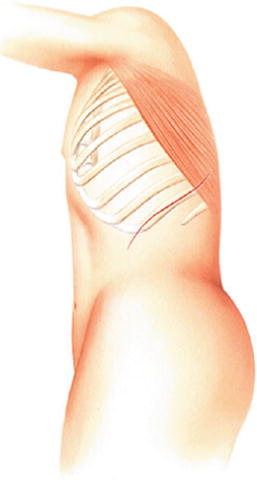
Fig. 21.1
A curved oblique incision is made along the 11th rib
2.
Along this incision line, m. latissimus dorsi is undermined and the muscle belly is clamped with two Pean’s or Kelly’s forceps in parallel. Between those two clamps, the muscle belly is incised using electric cautery. In the same manner, the underlying m. serratus anterior is incised (Fig. 21.2).
Fig. 21.2
Excision of m. latissimus dorsi and m. serratus anterior along the 11th rib
3.
Underneath those separated muscle layers, the 11th rib appears and the superficial periosteum is opened. The superior surface, then the inferior surface, followed by the undersurface of the 11th rib are carefully released subperiosteally using periosteal elevators such as Cobb and Dwyer instruments. Then, the 11th rib is harvested from the angle of the rib to the costal cartilage junction for bone grafting, leaving the 11th costal cartilage as the entrance to both the retroperitoneal and the extrapleural spaces. This cartilage, which forms a part of the arcus costalis, is then split with a sharp knife. The proximal stump of the 11th rib is packed with bone wax for hemostasis (Fig. 21.3).
Fig. 21.3
After removal of the 11th rib. The bed of the 11 th rib is made of the inner periosteum, the endothoracic fascia, and the parietal pleura. The interspace of the split costal cartilage, which is the entry zone to the retroperitoneal and the extrapleural spaces, is shown
Caution: The rib bed of the 11th rib is composed of three layers; the inner periosteum of the 11th rib, endothoracic fascia, and the parietal pleural. The rib bed should be maintained intact so as not to converge to thoracotomy.
4.
Because the diaphragm inserts to the 11th and the 12th costal cartilage in the lateral aspect of the thoracic cage, immediately after splitting the 11th costal cartilage, muscle fibers of the diaphragm spontaneously separate about 2 cm along its fibrous orientation. When both sides of the split cartilage are retracted, the m. obliquus externus abdominis is easily separated along its fibrous orientation, and the underlying m. obliquus internus abdominis and m. transversus abdominis appear. Both the m. obliquus interims abdominis and the m. transversus abdominis are carefully undermined and detached from the underlying peritoneum and then clamped with two Pean’s or Kelly’s forceps in parallel. Between those two forceps, muscle bellies are incised with electric cautery 3 to 5 cm along the incision line, until the underlying thin peritoneum becomes visible. M. transversus abdominis inserts to the arcus costalis and is interdigitated with insertion of the diaphragm (Fig. 21.4).