(1)
Marina del Rey, CA, USA
Deceased
For female patients, and for males of slender build, the transaxillary transthoracic approach is an alternative to the posterior parascapular approach to the upper dorsal spine. The highest dorsal vertebra reached through this approach is the body of T1, although the C7–T1 interspace is often accessible at the extreme apex of the thoracic inlet. The most significant advantage of this approach is that no major muscle groups are sectioned during the procedure, and there is rapid functional recovery of shoulder girdle mobility. The principal disadvantage of the approach is its limited exposure compared with that obtained by complete mobilization of the scapula. The approach is not indicated in obese individuals or in males of substantial muscular build with hypertrophic pectoralis major and latissimus dorsi muscles. The approach described here is from the right side, although the left-side approach is commonly used.
Get Clinical Tree app for offline access

1.
Position the patient with the right side of the torso elevated 60 degrees or more. The complete left lateral decubitus position can be used. Carefully protect the wrist and forearm to the elbow with padded circular dressings. Abduct the upper extremity and flex the elbow. Affix the padded forearm to an “L” bar at the anesthesiologist’s end of the table. Do not cover the arm with a sterile stockinette, as the operative field can be satisfactorily prepped and draped using standard orthopedic technique, excluding the portion of the upper extremity distal to the midbiceps (Fig. 15.1).
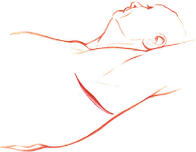
Fig. 15.1
The transaxillary approach to the upper dorsal spine. Position the patient with the left side of the torso elevated 60 degrees or more after protecting the wrist and forearms to the elbow with padded circular dressings. Abduct the upper extremity and flex the elbow. Drape the portion of the upper extremity distal to the midbiceps. The incision is transverse at the base of the natural axillary hairline
2.
Place the incision transversely at the base of the natural axillary hairline, and deepen it by electrocautery in the most direct manner to the chest wall. Divide the pectoralis musculature (Fig. 15.2).
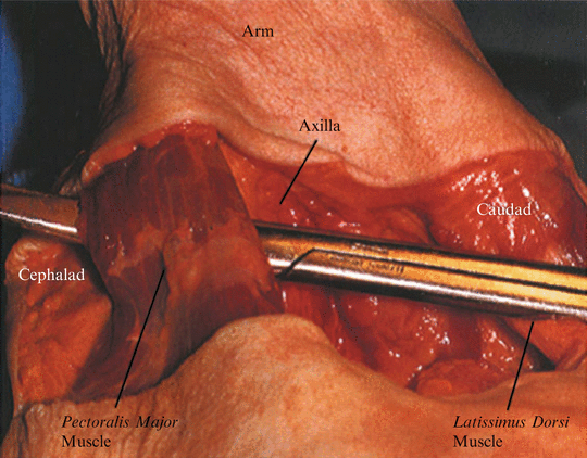
Fig. 15.2
Division of the pectoral muscles is not debilitating and responds to postoperative rehabilitation. Avoid tangential dissection into the succulent axillary tissue and cut directly to the chest wall
Caution: It is important to avoid undermining the succulent axillary soft tissues by oblique dissection.
3.
Once the chest wall is reached, dissect upward to the apex of the thorax in an avascular plane. By palpation, identify the tight circle of the 1st rib within the circle of the 2nd rib. The 3rd rib is of principal surgical interest for gaining access to the upper dorsal spine.
4.
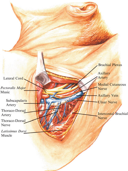
The relatively large sensory intercostobrachial nerve emerges from the second intercostal space and contributes innervation to the inner aspect of the entire upper arm, as well as a minor sensory “twig” that penetrates he pectoralis major muscle and innervates a patch of prepectoral skin (Figs. 15.3 15.4). Avulse this sensory nerve at its point of emergence from the intercostal space and excise a 4- to 5-cm peripheral segment of the nerve. Of course, you should inform the patient preoperatively that this procedure will be done and that there will be postoperative numbness of the denervated area. The altered sensation that follows surgery is not bothersome to most patients and is gradually overcome within several months by sensory ingrowth from adjacent neural segments.