Type of cyst
Location
Compression of the nervous structures
Hydrocephalus
Indication to transcranial surgery
Transcranial approaches
Site of fenestration and CSF drainage
Intrasellar
Sella turcica with suprasellar extension
None or slight chiasmal compression
No
Suprasellar extension, failed endoscopy
Supraorbital, subfrontal, pterional
Chiasmatic cistern
Suprasellar
Suprasellar cistern
Optic chiasm, hypothalamus, high brainstem, third ventricle
90 %
No hydrocephalus, failed endoscopy
Supraorbital, subfrontal, pterional, transcallosal
Basal cisterns
Interpeduncular
Interpeduncular cistern
Optic chiasm, hypothalamus, third cranial nerve
No or slight
No hydrocephalus, small cysts
Pterional
Chiasmatic and lamina terminalis cisterns
Retroclival
Prepontine cistern
Optic chiasm and tracts, pons and mesencephalon, fifth to eighth cranial nerves
Often present
Treatment of choice
Suboccipital retrosigmoid
Cerebellopontine angle and prepontine cisterns
Craniocervical
Anterior prebulbo-spinal
Posterior retrobulbospinal
Medulla, high spinal cord, lower cranial nerves, C1–C2 roots
No
Treatment of choice
Midline suboccipital
Prebulbar cistern, cisterna magna, and spinal canal

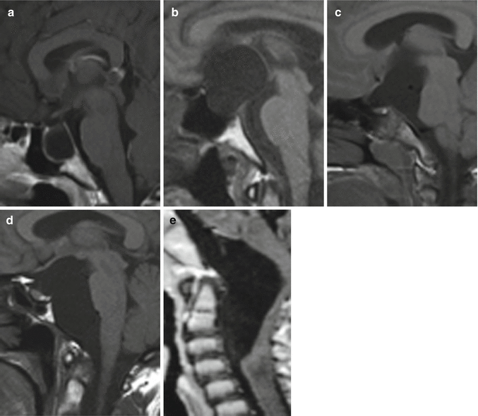
Fig. 17.1
Magnetic Resonance (MR), sagittal T1-sequences, of the various midline basal arachnoid cysts: (a) intrasellar; (b) suprasellar; (c) interpeduncular; (d) retroclival; (e) craniocervical (anterior)
1.
Intrasellar
2.
Suprasellar
3.
Interpeduncular
4.
Retroclival
5.
Craniocervical
1.
The intrasellar cysts lie within an enlarged sella turcica and may extend in the suprasellar cistern. They may show variable size and may become very large. Compression of the optic chiasm results in variable impairment of the visual function and bitemporal hemianopia [1]. Endocrinological disturbances from compression of the pituitary gland and stalk and hypothalamus often occur [2].
2.
The suprasellar cysts arise from intra-arachnoid dilatation and upward herniation of the superior (or mesencephalic) layer of the Liliequist’s membrane caused by cerebrospinal fluid (CSF) pulsation in the prepontine cistern [3]. Thus, they lie anterior to the interpeduncular cistern [4]. The third ventricle is compressed and displaced upwards, resulting in hydrocephalus, whereas the basilar bifurcation is pushed posteriorly against the brainstem. The suprasellar cysts may often be asymptomatic or may present with macrocephaly, intracranial hypertension syndrome, developmental delay, endocrine deficits, reduced visual field, and decreased visual acuity.
3.
The interpeduncular cysts lie within the interpeduncular cistern, between the diencephalic and mesencephalic leaves of the Liliequist’s membrane. Although these cysts have been considered a posterior variant of the suprasellar cysts [4], they are a peculiar, although rare, entity [5]. Interpeduncular cysts are small and tend to preserve the floor of the third ventricle, resulting in absence of hydrocephalus. Their limited expansion minimizes the effect on the neighboring structures (chiasm, CSF spaces). In the reported cases, the cyst was incidental or presented with third cranial nerve deficit [5–7]. Pure interpeduncular cysts tend to remain stable over time.
4.
The retroclival cysts arise posterior to the mesencephalic leaf of the Liliequist’s membrane and lie in the prepontine cistern. They are exceptional, with only several reported cases [8–13]. They may extend upward in the interpeduncular cistern and laterally to the cerebellopontine angle. The typical magnetic resonance features of retroclival cysts are vertical displacement of the optic chiasm and tracts, upward deflection of the rostral mesencephalon and mammillary bodies, and effacement of the ventral pons.
5.
The cysts of the craniovertebral junction are rare and may lie posterior or anterior to the bulbo-medullary junction. Anterior cysts are exceptional, with only a few reported cases [14, 15]. They lie in relationship with the clivus and C1 to C3 bodies; they compress and displace backward the medulla and high spinal cord and may stretch the lower cranial nerves and C1 to C3 roots. There is no hydrocephalus. These cysts may be asymptomatic [15] or may present with lower limb weakness and gait disturbances [14].
17.2 Management Options
The management options for the skull base midline arachnoid cysts include observation, endoscopy, shunting, and transcranial surgery.
Factors involved in the management decision include patient age, size and location of the cyst, and neurological symptoms.
The conservative management with follow-up magnetic resonance studies is indicated for incidental cysts with no neurological symptoms. However, large asymptomatic cysts with mass effect should be treated, particularly in children [20], and if they show progressive enlargement on serial magnetic resonance studies.
The endoscopic fenestration into the ventricles and cisterns is the best option for midline cysts of middle or large size, mainly with hydrocephalus; in such cases it offers the best chances for definitive treatment and avoids the risk of the craniotomy and shunt dependence [20–22]. However, the endoscopic approach may be difficult for not large cysts without hydrocephalus [22].
The cyst–peritoneal shunt is an effective and not invasive procedure for the complete obliteration of large cysts. However, it may also present several complications, including shunt failure, malfunction and infection, hemorrhage, and lifelong shunt dependence [22–24]. Moreover, the shunt procedure may be difficult in cases of not large midline basal cysts [22].
The microsurgical approach by craniotomy allows to largely fenestrate the cyst into the subarachnoid spaces and leaves the patient shunt independent. However, it is considered too aggressive for a usually scarcely symptomatic cyst. Besides, it carries not negligible operative morbidity and the risk of complications, such as damage of the neural structures, infection, and hemorrhage [22, 25]. Thus, less invasive and even keyhole approaches should be preferred.
Large opening and partial excision of the cyst wall are the aim of the transcranial surgery; on the contrary, complete removal of the cyst is neither possible nor necessary. However, cyst recurrence even after large fenestrations has also been reported [22].
17.3 Indications to the Transcranial Surgery (Table 17.1)
17.3.1 Intrasellar Cysts
17.3.2 Suprasellar Cysts
The endoscopic fenestration by ventricle-cyst-cisternostomy is the treatment of first choice for suprasellar arachnoid cysts and results in clinical remission and decrease of the cyst size in most cases [20, 21, 26–28]. However, endoscopy is impractical in presence of tiny ventricles. Besides, even repeated endoscopic procedures may fail. In these instances, open microsurgical fenestration of the cyst into the basal cisterns is a safe and effective option, which should be preferred to shunt insertion [21, 29]. If the cyst is huge and causes severe intracranial hypertension, the open fenestration allows immediate relief of the pressure and reduces the cyst [22, 29].
Stay updated, free articles. Join our Telegram channel

Full access? Get Clinical Tree


