Transient Loss of Consciousness: Seizures and Syncope


Transient Loss of Consciousness: Seizures and Syncope


Maromi Nei

Reginald T. Ho


Transient loss of consciousness in the elderly often presents one of the most difficult diagnostic challenges, particularly because of the high incidence of chronic medical conditions and associated medication usage and the fact that so many conditions may lead to loss of consciousness.

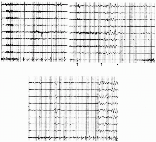
The major differential diagnoses include neurologic and cardiovascular causes, with seizures and syncope leading the list. Seizures, which often present in the elderly, frequently are related to vascular and neurodegenerative conditions. Syncope refers to transient loss of consciousness often accompanied by loss of postural tone and generally results from inadequate global cerebral nutrient perfusion (34). Sudden cessation of cerebral perfusion for only 6 to 8 seconds can cause syncope and diffuse slowing on an electroencephalogram (EEG) (Fig. 7-1) (48). Its incidence increases with age due in part to greater medication
usage and cardiovascular disease in elderly individuals (37,50,52).


Figure 7-1. The electrocardiogram (ECG) reveals asystole for approximately 15.5 seconds, beginning at the bold arrow. Diffuse slowing of the electroencephalogram (EEG) follows 6 seconds later, and suppression of cerebral activity follows 10 seconds later. The suppression continues for approximately 18 seconds, including 12 seconds after return of the cardiac rhythm. This is again followed by diffuse slowing of the EEG, which is later followed by return of the EEG to a normal waking pattern (not shown).


PRESENTATION AND DIFFERENTIAL DIAGNOSIS

Details regarding the symptoms preceding and following the event are critical to determining the cause for loss of consciousness (8,32,35). A history regarding specific symptoms preceding loss of consciousness, particularly the duration and quality, should be elicited, and any triggering events should be identified (Tables 7-1 and 7-2).


SEIZURES

Seizures generally present as stereotyped spells that follow a specific and consistent progression of symptoms during each event. In partial-onset seizures, a specific aura can occur before the onset of alteration in level of consciousness. A clue may be a specific aura occurring in isolation as well as at the onset of a complex partial seizure (those associated with alteration in level of consciousness but without generalization) or secondarily as a generalized tonic-clonic seizure. The duration of seizures is brief, generally less than 2 minutes. However, in seizures associated with an alteration in level of consciousness, patients often experience amnesia of the seizure itself and may not recall events immediately preceding or following the seizure. Postictal confusion can last for several minutes to hours.


Table 7-1. Spells in the Elderly: Differential Diagnosis


Neurologic


Seizure


Complex partial


Secondarily generalized tonic-clonic


Absence


Nonconvulsive status epilepticus


Transient ischemic attack


Basilar artery ischemia


Transient global amnesia


Migraine


Sleep disorder


Nonepileptic psychogenic seizure


Cardiac


Obstruction to outflow (aortic stenosis, idiopathic hypertrophic subaortic stenosis, pulmonary embolus)


Loss of effective pump function (myocardial infarction, tamponade)


Arrhythmias (bradyarrhythmias, tachyarrhythmias)


Reflexogenic


Vasovagal syncope


Situational syncope (cough, micturition, swallowing)


Carotid sinus hypersensitivity


Orthostatic hypotension


Medication effect


Hypovolemic


Neurogenic


Metabolic


Hypoglycemia


Hyperventilation


FOCAL CEREBRAL ISCHEMIA

Focal cerebral ischemia resulting in transient alteration in level of consciousness is not common. Normal consciousness depends on the functioning of both cerebral hemispheres, the reticular formation, other upper brainstem structures, the thalamus, and the hypothalamus (44). Thus, focal cerebral ischemia, as during a transient ischemic attack or stroke, must involve either both cerebral hemispheres or the brainstem and other deeper structures to result in alteration in level of consciousness. Posterior circulation ischemia or massive hemisphere infarction with shift can present with alteration in level of consciousness. Posterior circulation ischemia generally results in focal signs and symptoms (e.g., diplopia, eye movement abnormalities, other cranial nerve abnormalities, cerebellar dysfunction, motor and sensory dysfunction), which aid in the diagnosis. Massive infarction, of course, results in a sustained alteration in level of consciousness.


TRANSIENT GLOBAL AMNESIA

Transient global amnesia (TGA) presents with marked anterograde amnesia that generally persists for hours, as well as retrograde amnesia. Although patients may be disoriented to time and place, they retain knowledge of their identity. The patient often repeatedly asks the same questions and has difficulty encoding new memories during this event. No focal neurologic deficits are seen, and the patient is fully conscious throughout the episode, unlike during a complex partial seizure. The pathophysiology is debated but may be related to either cerebral ischemia or seizure. The finding of reversible changes in the CA-1 sector of the hippocampus on high-resolution magnetic resonance imaging (MRI) suggests that this area may be involved in the pathophysiology of TGA (6). The incidence of TGA has been estimated to be approximately five in 100,000 persons. Less than 25% of patients experience recurrent episodes (2,41).


SLEEP DISORDERS

Sleep disorders (e.g., sleep apnea or narcolepsy) present with other symptoms that suggest a sleep
disorder, particularly excessive daytime sleepiness, which results in lapses in consciousness. However, a history of sedation and concomitant symptoms (e.g., snoring or apnea) clearly differentiates the diagnosis of sleep disorders from other disorders.


Table 7-2. Variables That Distinguish Common Spells in the Elderly


Variable


Seizure


Syncope


TIA


TGA


Premonitory symptoms


None vs. aura


None vs. N/V, lightheadedness


None


None


Diaphoresis


Posture effect


None


Often erect


None


None


Onset


Acute


Variable


Acute


Acute


Bystander observations


Duration


1-2 minutes


Seconds to minutes


Minutes to hours


Hours


Movements


Variable tonic-clonic movements


Loss of tone Clonic jerks


Deficits along vascular pattern


None


lncontinence


Variable


None


None


None


Heart rate


Increased or decreased


Variable


Normal


Normal


Electroencephalogram during ictus


Epileptiform pattern


Diffuse Slowing


Focal Slowing or normal


Rare Slowing


Trauma


Tongue laceration or ecchymoses


Ecchymoses or fracture


None


None


Offset


Confusion Sleep


Alert or mild confusion


Alert


Alert


N/V, nausea and vomiting; TGA, transient global amnesia; TIA, transient ischemic attack.


NONEPILEPTIC PSYCHOGENIC SEIZURES

Nonepileptic psychogenic seizures are more common in younger individuals; however, they also occur in the elderly. A variety of different types of presentations are seen. Although slumping and sudden apparent loss of consciousness can occur, shaking and other movements can occur as well. Common features include (a) nonstereotyped spells; (b) irregular, nonrhythmic movements; (c) eye closure during the event; (d) waxing and waning of symptoms; (e) prolonged symptoms over several minutes to hours; (f) no history of spells arising directly from sleep; and (g) no history of severe injury (e.g., fracture, burn) during the spells. The patient may have a history of sexual or physical abuse, lack of response to anticonvulsant medication, and history of a psychiatric disorder. Recent data suggest that, in the elderly, a history of severe physical health problems or health-related traumatic experiences may be a prominent risk factor in this age group (20). Video-electroencephalographic (VEEG) monitoring (1) or EEG and observation of the episode are helpful in establishing the diagnosis.


SYNCOPE

Because syncope has a myriad of etiologies, it is useful to classify its causes into (a) cardiovascular, (b) neurally mediated (reflexogenic), (c) orthostatic (postural), and (d) metabolic because the diagnostic evaluation and prognosis in each category differ. Cardiovascular causes of syncope include arrhythmias and structural cardiopulmonary disease, and they should be considered in any elderly patient with significant heart disease (e.g., myocardial infarction, congestive heart failure). Arrhythmic syncope is often abrupt, and such an episode in a patient with left ventricular dysfunction or conduction abnormalities (e.g., bundle branch block) should raise suspicion for ventricular tachyarrhythmias and bradyarrhythmias, respectively. Structural cardiopulmonary disease causing syncope reduces cerebral perfusion by obstructing blood flow (e.g., aortic stenosis, hypertrophic obstructive cardiomyopathy) or decreasing cardiac output (e.g., myocardial infarction, tamponade).

Neurally mediated syncope includes vasovagal, viscerovagal (situational), and carotid sinus hypersensitivity and is due to an exaggerated reflex that increases vagal tone (causing bradycardia) while reducing sympathetic outflow (causing hypotension). Such forms of syncope often have a triggering event (e.g., prolonged standing, defecation, coughing). The classical prodromal symptoms of vasovagal syncope (warmth, nausea, lightheadedness, and diaphoresis) might be absent in
an elderly patient. Abrupt syncope without prodrome can even occur (malignant vasovagal syncope). Orthostatic syncope is due to an abrupt drop in blood pressure while assuming an erect posture and is common in the elderly (54). Autonomic dysfunction, loss of baroreceptor responses, and frequent use of multiple medications predispose elderly individuals to orthostatic syncope. Primary autonomic failure can be caused by multiple system atrophy, which is generally associated with brainstem dysfunction or parkinsonism. The Shy-Drager and Bradbury-Eggleston types of autonomic failure are associated with other evidence of autonomic dysfunction, including sexual and bladder dysfunction and anhidrosis. Secondary autonomic dysfunction can result from an autonomic neuropathy, often associated with a peripheral neuropathy, which may be seen in diabetes mellitus, chronic inflammatory demyelinating neuropathy, amyloidosis, and other types of neuropathy. Metabolic abnormalities causing syncope are rare. These include high-altitude sickness [causing low partial pressure of oxygen (PO2)], acute hyperventilation [causing low partial pressure of carbon dioxide (PCO2) and cerebral vasoconstriction], and hypoglycemia. Insulinomas can cause repetitive seizurelike spells due to recurrent hypoglycemia (7). Hypoglycemia is also often associated with neuro-adrenergic symptoms (e.g., diaphoresis and tremors).


SEIZURES

Seizures are among the most common causes of transient loss of consciousness in the elderly. The incidence of both acute symptomatic and unprovoked seizures increases with age. The incidence of acute symptomatic seizures is approximately 100 in 100,000 persons older than 60 years of age (5,39). These seizures may be caused by acute cerebrovascular ischemia (40% to 50%), metabolic derangements (10% to 15%), drug withdrawal, central nervous system (CNS) infection, acute trauma, or toxic insults (5,39). The incidence of unprovoked seizures exceeds 100 in 100,000 persons in this same age group (25). Unprovoked seizures can occur as the result of a prior stroke or head trauma or in association with degenerative diseases such as Alzheimer’s disease. However, a definitive cause for epilepsy (having two or more unprovoked seizures) is identified only in the minority of patients (30% to 50%) (5,39).

Risk factors for the development of epilepsy include a history of stroke, which is associated with a more than 20-fold increase in the risk of epilepsy; brain neoplasm; neurodegenerative disease; drug or alcohol withdrawal; CNS infection, which is associated with a threefold increase in risk; and head trauma, which is associated with a threefold increase in risk (33,49). Alzheimer’s disease is associated with a fiveto 10-fold increase in risk for epilepsy (18,27,29,40,46). Major depression may also be a risk factor for seizures in the elderly (30).

Multiple seizure types may be seen. Partial-onset seizures, often related to degenerative, vascular, or neoplastic causes, are most common. However, generalized seizures related to toxic-metabolic encephalopathies and, perhaps, to genetic predisposition also occur.


TEMPORAL LOBE SEIZURES

Temporal lobe seizures are the most common type of seizure in the adult population, although extratemporal seizures may be more common in the elderly than in other age groups. Temporal lobe seizures can begin with an aura, such as a feeling of epigastric discomfort, an indescribable sensation, déjà vu, fear, or tinnitus. Although seizures are typically associated with positive phenomena, rather than the absence of normal function, both types of symptoms can be seen. During the seizure, speech arrest can occur, particularly in seizures originating in the left hemisphere. Complex partial seizures (those associated with an alteration in level of consciousness) arising from the temporal lobe may be manifested by automatisms, lip smacking, teeth grinding, chewing, and the utterance of phrases or sentences that can be unintelligible or repetitive. Staring without automatisms is a common presentation in this age group, and head or eye deviation or dystonic posturing can occur. Postictally, confusion can last for several minutes to hours, and the patient may experience amnesia of the seizure. Because the temporal lobes are important for memory, patients with seizures arising from this area often complain of memory difficulty interictally as well.

Because of connections to the hypothalamus, temporal lobe seizures can be associated with autonomic changes. These include pupillary dilatation, apnea, hyperventilation, flushing, diaphoresis, urinary urgency or incontinence, and heart rate and rhythm changes. Both ictal tachycardia and bradycardia can occur during seizures arising from the temporal lobes. Most temporal lobe seizures are associated with an increase in heart rate, but supraventricular tachycardia, sinus arrest, atrial fibrillation, and frequent premature atrial and ventricular depolarizations can occur (11,43). This association is particularly important to keep in mind when interpreting electrocardiographic (ECG) telemetry data during a typical spell. The ECG data alone without concomitant EEG during the spell may suggest that the event is primarily arrhythmic in origin, when in reality, the seizure is the primary event. Thus, simultaneous EEG-ECG monitoring is important in these cases.

Only gold members can continue reading. Log In or Register to continue

Stay updated, free articles. Join our Telegram channel

Jul 14, 2016 | Posted by in NEUROLOGY | Comments Off on Transient Loss of Consciousness: Seizures and Syncope

Full access? Get Clinical Tree

Get Clinical Tree app for offline access