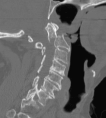
2 A 63-year-old woman fell while riding a bike. She sustained a type II odontoid fracture and was initially treated with halo immobilization for 6 weeks. On follow-up x-ray, she continued to have subluxation but remained neurologically intact. FIGURE 2-1 Reconstructed CT of the C-spine shows a prominent subluxation between the fracture fragment and body of the dens. Computed tomography (CT) reconstruction of the C-spine (Fig. 2-1) is notable for a type II odontoid fracture with severe subluxation of over 10 mm. FIGURE 2-2 Postoperative x-ray with odontoid screw in place. Failure of orthosis treatment of type II odontoid fracture An odontoid screw was placed (Fig. 2-2). Optimum reduction was achieved with this fixation. Odontoid fractures are usually an extension injury. Type I odontoid fractures involve the tip of the odontoid and are stable. They can usually be treated by a cervical collar. Type II fractures are the most common, and the fracture site is at the junction of the odontoid peg and the C2 body. These fractures need either halo immobilization or operative fixation. Type III fractures involve the body of C2, and while some physicians feel union may occur in a collar, others advocate operative intervention. Surgical treatment for type II odontoid fractures is recommended if there is over 6 mm translation, comminuted fragment (type IIa), or C2-C3 in anterolisthesis over 3 mm. The question arises whether to fixate a type II odontoid fracture anteriorly or posteriorly. Criteria for anterior fixation through an odontoid screw include sufficient neck extension ability, an intact transverse ligament, a non-barrel chest, and an ability to line up the fractured bone. This technique fixes the fracture directly without bone graft and avoids loss of rotational motion in comparison to posterior techniques. Poor bone quality, transverse ligament failure, an inability to reduce the fracture, a fracture line through the intended screw course, and chronic nonunion are contraindications to odontoid screw placement. Posterior fixation through transarticular screws carries higher neurovascular morbidity but provides more rigid arthrodesis. In addition, this technique is more useful in atypical type II fractures, C1-C2 fracture dislocations (intraoperative reduction is obtained), and cases that need decompression. Patients with significant thoracic kyphosis are notoriously difficult to fixate through transarticular screws. Again, the patient’s spine and body anatomy will dictate the best approach. Borm W, Kast E, Richter HP, Mohr K. Anterior screw fixation in type II odontoid fractures: is there a difference in outcome between age groups? Neurosurgery 2003;52:1089–1092
Transodontoid Screw for Type II Odontoid Fracture
Presentation
Radiologic Findings

Diagnosis
Treatment
Discussion
SUGGESTED READING
< div class='tao-gold-member'>
Transodontoid Screw for Type II Odontoid Fracture
Only gold members can continue reading. Log In or Register to continue


Full access? Get Clinical Tree


