(1)
Marina Spine Center, Marina del Rey, CA, USA
1.
Position the patient supine over the flexion crease in the table and hyperextend the lumbar spine for greater exposure.
2.
Make either a vertical midline incision or the transverse “smile” incision. The “smile” is better cosmetically and gives excellent exposure but requires transection of the rectus abdominis muscle (Figs. 29.1, 29.2). Identify the rectus sheath; open the anterior portion of the rectus sheath. Transect the rectus abdominis muscle. The posterior rectus sheath, the abdominal fascia, and the peritoneum are conjoined in this area. Incise carefully through the posterior rectus sheath and abdominal fascia to the peritoneum (Fig. 29.3).
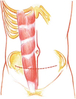
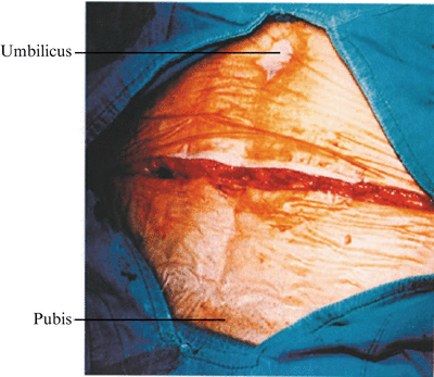
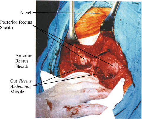
Fig. 29.1
Position the patient supine over the flexion crease in the table and hyperextend the lumbar spine for greater exposure. Make a vertical midline incision or the transverse “smile” incision. The smile incision is better cosmetically, gives excellent exposure, and requires transection of the rectus abdominis muscle
Fig. 29.2
Transverse “smile” incision
Fig. 29.3
Open the anterior portion of the rectus sheath and transect each rectus abdominis muscle. Identify the posterior rectus sheath. The posterior rectus sheath, abdominal fascia, and peritoneum are conjoined in this area
3.
Pick up the peritoneum (Fig. 29.4). Open it carefully to avoid bowel damage and extend the opening the length of the wound. Pack off the bowel and identify the posterior peritoneum over the sacral promontory.
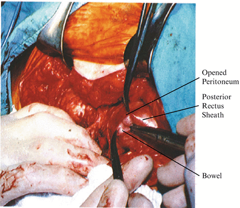
Fig. 29.4
Incise carefully through the posterior rectus sheath and abdominal fascia and identify the peritoneum. Pick up the peritoneum, carefully open the peritoneum to avoid bowel damage, and extend the peritoneal opening the entire length of the wound. Insert a self-retaining Deaver-type retractor
4.
Get Clinical Tree app for offline access

Palpate the aorta and both iliac vessels through the posterior peritoneum (Figs. 29.5, 29.6). Identify the softer texture of the L5–S1 disc.
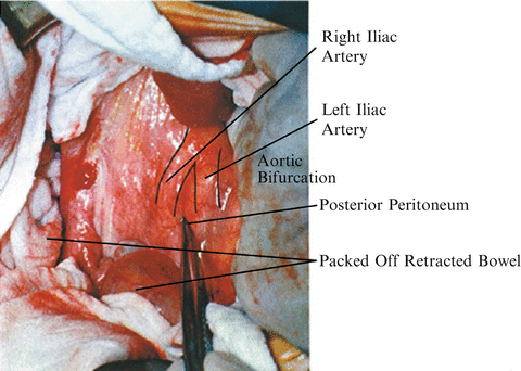
Fig. 29.5


Pack off the bowel to allow adequate exposure of the posterior peritoneum over the sacral prominence. Palpate for the aortic pulse and common iliac artery. With identification of the sacral prominence and the bifurcation of the aorta, one may inject the posterior retroperitoneum with saline to allow elevation of the peritoneum off the important vascular structures
Related posts:


Stay updated, free articles. Join our Telegram channel
Full access? Get Clinical Tree