, Alfred A. Steinberger1 and Frank Moore1
(1)
Mount Sinai Hospital, New York, NY, USA
The cervicothoracic junction represents one of the more complex areas of the spine because of the proximity of visceral and vascular structures at the thoracic inlet. With the introduction of magnetic resonance imaging (MRI), it is apparent that most tumors, infections, and degenerative processes result in compression of the cord from a ventral direction. Logically, therefore, direct anterior approaches offer the best exposure for direct decompression of the spinal cord. In the majority of cases, restoration of spinal stability by reconstruction of the resected segments represents an equally important goal of surgery. Such reconstruction should be carried out in conjunction with instrumentation to achieve rigid and immediate internal fixation.
In 1957, Cauchoix and Binet described a direct approach to the cervicothoracic region through median sternotomy.1 In their classic approach, the skin incision included two parts: a cervical component along the anterior border of the sternomastoid muscle and a thoracic incision in the midline, carried down to the xyphoid process. The sternum was completely split and retracted. Although median sternotomy provides complete access to the cervicothoracic segments from C4 down to T4, complete median sternotomy is not always indicated for standard approaches. During the past decade, we and others have described a variety of modifications to this original operative procedure [2 – 8].
Clinical indications for this operative exposure include not only lesions involving the spine but also tumors of the superior mediastinum (thymomas), as well as the management of vascular injuries involving the innominate artery [7 – 9]. For spinal lesions, this approach is indicated for tumors, tuberculosis involving the cervicothoracic junction, trauma with collapse of the upper thoracic vertebra, and the correction of cervicothoracic kyphosis.
Surgical Anatomy
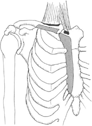
The thoracic inlet is kidney shaped with an average anteroposterior diameter of 5 cm and a transverse diameter of 10 cm. It is bounded by the first thoracic vertebra posteriorly, the top of the manubrium anteriorly, and the 1st ribs on each side. Its plane slopes forward and downward, but there is considerable variation in the size, shape, and obliquity of the inlet. The sternomastoid muscle arises from the sternum and clavicle by two heads (Fig. 14.1). The sternal head is a rounded fasciculus that is tendonous and arises from the upper part of the anterior surface of the manubrium. The clavicular head is more fleshy and arises from the superior border and anterior surface of the medial third of the clavicle. The infrahyoid strap muscles lie more posteriorly. The sternohyoid muscle arises from the posterior surface of the medial end of the clavicle, the posterior sternoclavicular ligament, and the posterior surface of the manubrium and medial end of the 1st rib. Important vascular structures in the superior mediastinum include the innominate, common carotid, and left subclavian arteries arising from the arch of the aorta. The innominate artery originates from the convexity of the aortic arch and passes obliquely upward, backward, and to the right. A high-riding arch with a more distal origin of the innominate artery may necessitate considerable retraction if a right-sided approach is used.


Fig. 14.1
Muscular and tendonous attachment of the origin of the sternocleidomastoid muscle is shown on the sternum and the clavicle. The sternal head is tendonous and arises from the anterior superior part of the manubrium. (From O’Shea J, Sun-daresan N, Stein Berger A, Moore F. In Menezes A, Sonntag VKH (eds): Principles of Spinal Surgery. New York, McGraw-Hill, 1996, p 1254, with permission.)
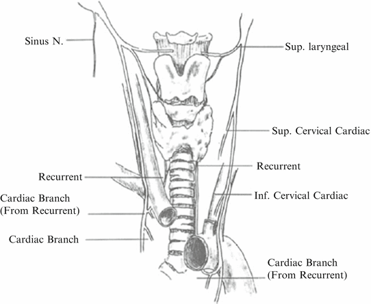
The vagus nerve and its branches are the most important nerves to identify in the neck (Fig. 14.2). The vagus nerve is (1) motor to all smooth muscle, (2) secretory to all glands, and (3) afferent from all mucous surfaces in the following parts: the pharynx (lowest part), larynx, trachea, bronchi, and lungs; esophagus (entire), stomach, and gut down to the left colic flexure; liver, gallbladder, and bile passages; pancreas and pancreatic ducts; and perhaps spleen and kidney; (4) motor to all muscles of the larynx, all muscles of the pharynx (except the stylopharyngeus), and all the muscles of the palate (except the tensor veli palatini); (5) the conveyor of taste from the few taste buds about the epiglottis; (6) inhibitory to cardiac muscle; and (7) sensory to the outer surface of the eardrum, the external acoustic meatus, and the back of the auricle. In the neck, the vagus nerve gives (1) a pharyngeal branch to the superior and middle constrictors and muscles of the soft palate; (2) the superior laryngeal nerve, via the internal laryngeal nerve, is sensory to the larynx above the vocal cords and to the lowest part of the pharynx and, via the external laryngeal nerve, motor to the inferior constrictor and cricothyroid and (3) a twig (sinus nerve) to the carotid sinus, and (4) two cardiac branches.


Fig. 14.2
The branches of the vagus nerve in the neck include the recurrent layngeal nerve, the superior clavicular nerve, the superior laryngeal nerve, and inferior cervical cardiac and cardiac branches from the recurrent laryngeal nerve. (From O’Shea J, Sundaresan N, Stein Berger A, Moore F. In Menezes A, Sonntag VKH (eds): Principles of Spinal Surgery. New York, McGraw-Hill, 1996, p 1255, with permission.)
The recurrent laryngeal nerve arises from the vagus and courses around the subclavian artery on the right and around the aortic arch on the left. It traverses the operative field obliquely at a higher level on the right; on the left side, it reaches the tracheoesophageal groove more caudally and is thus less liable to injury with a left-sided exposure. In addition, the recurrent laryngeal nerve may occasionally be nonrecurrent on the right side and take a more direct course. On the left side, no such anatomical variations are seen. For this reason, a left-sided approach to the cervicothoracic region is generally recommended.
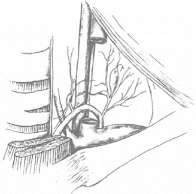
Another important structure that is potentially vulnerable to injury is the thoracic duct (Fig. 14.3). At this level, it usually lies to the left of the midline and empties at the junction of the internal jugular and subclavian veins posteriorly. Occasionally, it may divide into two branches, one emptying on the left side and the other into the right subclavian vein with the right lymphatic duct. The right lymphatic duct is much less prominent and should not be encountered during this dissection.


Fig. 14.3
The thoracic duct was to the left of the midline and empties into half the junction of the internal jugular subclavian veins posteriorly. (From O’Shea J, Sundaresan N, Stein Berger A, Moore F. In Menezes A, Sonntag VKH (eds): Principles of Spinal Surgery. New York, McGraw-Hill, 1996, p 1255, with permission.)
Operative Approach
The operation is performed under general endotracheal anesthesia with the patient placed in the supine position. In patients with unstable spines, awake nasotracheal intubation may be performed at the discretion of the surgeon. Arterial lines and wide-bore intravenous lines are used, as well as compression stockings for the lower extremities. The neck is extended slightly using a folded sheet under the shoulders and a doughnut under the head. The range of extension and flexion of the neck is tested preoperatively with the patient awake. In patients with obvious instability, a halo traction device is applied and traction maintained intraoperatively. We currently monitor somatosensory evoked potentials (SSEP) routinely, as well as an image intensifier that is draped in place. Both arms are positioned by the sides, and traction bands are applied to both wrists to pull the arms down for lateral radiographic imaging during the procedure.
1.
Get Clinical Tree app for offline access

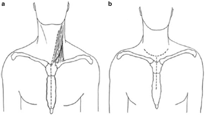
Two different skin incisions may be used: a vertical incision along the medial aspect of the sternomastoid extending along the midline of the sternum down to the xyphoid, or a transverse incision 1 cm above the clavicle that is then extended in the shape of a T over the sternum (Figs. 14.4a, 14.4b).


Fig. 14.4


(a) The vertical incision extends along the medial border of the sternocleidomastoid to the midline of the sternum extending down the xyphoid process. (b) The transverse incision is 1 cm above the clavicle and then is extended in the shape of a “T” over the sternum. (From O’Shea J, Sundaresan N, Stein Berger A, Moore F. In Menezes A, Sonntag VKH (eds): Principles of Spinal Surgery. New York, McGraw-Hill, 1996, p 1256, with permission.)
Stay updated, free articles. Join our Telegram channel

Full access? Get Clinical Tree


