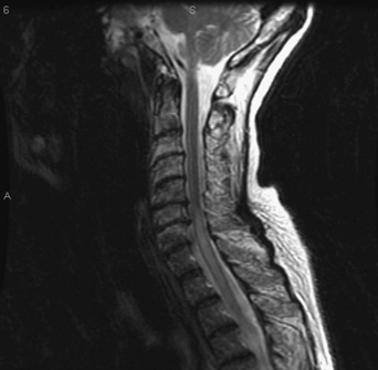
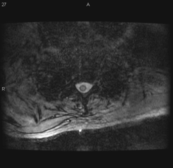
36 A 61-year-old woman with a history of autoimmune illness recently became progressively paraparetic with incontinence. She complained of a new-onset “band”-like feeling in her chest. On examination, she had a thoracic sensory level and was areflexic and flaccid in her lower extremities. Magnetic resonance imaging (MRI) of the thoracic spine reveals a hyperintense lesion that is intramedullary but not widening the cord (Figs. 36-1 and 36-2). MRI is the imaging test of choice in these cases. FIGURE 36-1 Sagittal MRI of the thoracic spine shows a midthoracic hyperintense lesion that is not widening the cord. Transverse myelitis While the patient was hospitalized, a cerebrospinal fluid (CSF) sample was sent for routine laboratory assessment as well as immunoglobulin G (IgG) index, oligoclonal bands, and flow cytometry. The results were inconclusive. Steroids were given, and the patient eventually underwent rehabilitation. Repeat imaging was undertaken at a later time.
Transverse Myelitis
Presentation
Radiologic Findings

Diagnosis
Treatment
| Degenerative |
| Metabolic |
| Trauma |
| Congenital |
| Vascular |
| Stroke, arteriovenous malformation or fistula, cavernous angioma, granulomatous angiitis |
| Demyelination |
| Multiple sclerosis |
| Neoplasm |
| Epidermoid, dermoid, lipoma |
| Hemangioblastoma |
| Ependymoma |
| Astrocytoma, ganglioglioma, xanthoastrocytoma, oligodendroglioma |
| Leptomeningeal gliomatosis |
| Intramedullary schwannoma |
| Metastasis |
| Lymphoma |
| Inflammatory/infection |
| AIDS, neurosyphilis, viral, vaccine, autoimmune |
| Parasitic: cysticercosis, sparganosis, schistosomiasis, toxoplasmosis |
| Granuloma |
| Sarcoid, tuberculosis, brucella, histoplasmosis, vasculitis |
| Vitamin deficiency |
| Systemic illness, drug related |
*Courtesy of Dr. Jack Rock, M.D.


