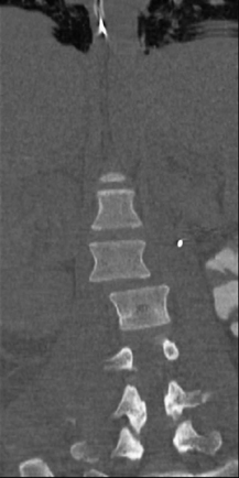
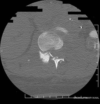
13 An 18-year-old woman was hit by a car and sustained multisystemic injury. On presentation she was paraplegic and had complete spinal cord injury in the lower extremities. She had a prolonged intensive care unit course and was finally stable enough for surgery. FIGURE 13-1 CT AP reconstruction of the thoracolumbar junction shows an L1–L2 fracture dislocation. Anteroposterior (AP) reconstruction of the thoracolumbar junction shows an L1–L2 fracture dislocation. An axial slice through the area demonstrates the rotational translation of L1 on L2 (Figs. 13-1 and 13-2). Fracture dislocation of the lumbar spine FIGURE 13-2 An axial CT slice through the area demonstrates the rotational translation of L1 on L2.
Traumatic Lumbar Fracture
Presentation
Radiologic Findings
Diagnosis
| Approach/Level | Ventral | Ventrolateral | Dorsolateral | Dorsal |
| C6-T3 | Cervicothoracic | — | Lat. parascapular extrapleural | Transpedicular Translaminar |
| T3-T10 | Transthoracic | Retropleural | Lat. extracavitary | Transpedicular Translaminar |
| T10-L5 | Thoracoabdominal | Retroperitoneal | Costotransversectomy | Transpedicular Translaminar |
Treatment
Decompression, reduction, and thoracolumbar pedicle screw fixation were undertaken through a posterior approach.
Discussion
Fracture dislocations of the spine are the result of severe forces that disrupt all three columns by combining rotation, tension, compression, and shear. Flexion rotation fractures often occur through the disc space. These are associated with abdominal injury and neurologic damage. Treatment focuses on realignment of the spinal column, posterior stabilization, and early mobilization.
The surgical approach of a thoracolumbar fracture depends on location, mechanism of injury, neurologic status, and overall clinical status. Table 13-1 summarizes the approaches.
SUGGESTED READING
Chaloupka R. Complete rotational burst fracture of the third lumbar vertebra managed by posterior surgery. A case report. Spine 1999;24:302–305
< div class='tao-gold-member'>


