(1)
Swingle Clinic, Vancouver, BC, Canada
Keywords
Neuroguided stimulationStress toleranceSeizure disordersAge-related declinesAnxietyDepressionBipolar disordersADHDSleep disordersEmotional traumaFamily dynamicsLeft handedSeniorsAs has been stated several times, neurotherapy blends perfectly with other treatment modalities and metaphors. Neurotherapy closes the gap in many therapies in that the clinician will be able to attend to the biological component of treatment. Neurotherapies, in any of the many manifestations and methodologies, including neurofeedback and neuroguided stimulation (braindriving), are NOT stand-alone treatments. They are simply not adequate, on their own, to treat any meaningful clinical condition. Further, if one is truly treating the condition and not the label, then any one-size-fits-all procedure is also simply inadequate.
One-size-fits-all neurotherapies do, however, come in many guises if not sizes. “Dynamical” models of treatment purport that if one gives the brain a good shake-up or workout, there will be a general reorganization tending toward normative, state-sturdy baselines. And like all notions about interconnected systems, there is some theoretical, if not empirical, merit to this position.
Serious scientist/practitioners working with database-guided therapies (e.g., z-score training) do show that interconnectivity among brain sites become more “normalized” with such treatments. We do know that improved connectivity is associated with client reports of improved functioning with traumatic brain-injured clients. So there may be some merit for including shake-the-pot sessions as part of neurotherapy packages for some clients. But, there is scant evidence for shake the pot as a one size fits all for all clinical clients. As stated previously, departures from normative functioning need not be associated with any clinical complaint. In fact, such departures from the “normal” may be associated with unique skills and approaches.
Most of the flagrantly bogus one-size-fits-all franchise-like enterprises feature some form of shake the pot. Some, however, are a bit more sophisticated and have specific shake-the-pot protocols for specific label complaints such as depression and ADHD. Symptom-based protocols are much too imprecise for reasons already stated. This lack of precision is apparent regardless of whether or not the base treatment metaphor is shake the pot or treating (activating or quieting) presumed brain regions associated with the condition or on general notions such as relaxation/quieting protocols benefit everyone.
However, with clinical populations, there are specific comprehensive neurotherapy protocols that can have general beneficial effects for many clients. These protocols have emerged from clinical practice and clinical databases. The efficacy of these general protocols is based on QEEG data indicating neurological commonalities among clinical clients.
General Protocols
Stress Tolerance
Increase the Theta/Beta ratio in the occipital region of the brain (sites O1 and O2). This protocol emerged from the early work of Gene Peniston (Peniston and Kulkosky 1999) who demonstrated that increasing the amplitude of Theta and Alpha at the back of the brain had substantial beneficial effects with chronic hospitalized alcoholics. The “Alpha/Theta” protocol has been found to have benefit with clients with other complaints as well.
The Theta/Beta ratio can be enhanced with several protocol variations. The original Alpha/Theta training protocol is to initially increase Alpha amplitude and then switch over to increasing Theta amplitude. Variations on this protocol include going directly to increasing Theta amplitude and/or decreasing Beta amplitude. The latter is particularly effective when the client’s Theta/Beta ratio is low because of elevated Beta and less the result of deficient Theta amplitude.
The reason that increasing the Theta/Beta ratio has general beneficial effects for many clients is because over 70 % of clinical clients (in our sample population) have a Theta/Beta deficiency in the occipital region of the brain. Further, that percentage increases to over 80 % for clients diagnosed with bipolar disorder. The “emergency” braindriving protocol, described in Chap. 6, includes Theta enhancement.
Clinicians must be mindful that increasing the Theta/Beta ratio can have the effect of “releasing” blunted Alpha which is related to emotional trauma. In our database, about 40 % of general clinical clients show a trauma marker, and this percentage jumps to a stunning 87 % with clients diagnosed with a bipolar disorder. Hence, client receiving this “relaxation” therapy may have an abreactive episode when the emotional content associated with traumatic events surface during the treatment. This emotional abreaction has been described by many clinicians who train up Alpha and/or Theta amplitude posteriorly.
Clinicians working with other forms of relaxation therapies have also commented on relaxation-induced panic episodes with clients. Clinically, of course, these releases are very therapeutically beneficial, and clinicians frequently use such procedures for precisely this purpose. However, these potential effects of such training further emphasize that only licensed healthcare professionals should be practicing in these arenas. Further, there are going to be some potentially harmful outcomes from the unlicensed practitioners who include this generally beneficial protocol into their one-size-fits-all programs.
Body Quiescence
Based on the seminal work of Barry Sterman (Sterman and Egner 2006; see also Robbins 2000) on the treatment of seizure disorders, protocols for increasing the sensory motor rhythm (13–15 Hz) amplitude over the sensory motor cortex (locations C3, Cz, and C4) have been shown to have a generally beneficial effect on decreasing physical arousal. Often this training includes simultaneous reduction in the amplitude of Theta as well. SMR training has been shown to improve sleep quality (Hoedlmoser et al. 2008), motor tics (Tansey 1986), hyperactivity (Lubar and Shouse 1977), and pseudoseizures (Swingle 1998).
Brain Brightening
Cognitive functioning is generally improved when the amplitude of slow-frequency EEG is reduced (cf. Abstra-Angelakis et al. 2007; Swingle 2002) over the frontal cortex or the sensory motor cortex. This can be accomplished in several ways including reducing the amplitude of EEG activity in the 3–9 Hz range or by increasing the peak frequency of Alpha (8–12 Hz). There are several variations in these brain-brightening protocols such as reducing Theta amplitude while simultaneously increasing Beta (16–25 Hz) amplitude. Some protocols focus on just increasing Beta amplitude, others on reducing Theta. Bandwidth of the slow frequency varies as well with some clinicians preferring to focus on Theta and others on low Alpha (8–9 Hz), and some prefer to train across the entire bandwidth (3–9 Hz). Decreasing Delta (1–3 Hz) likewise can have a cognitively brightening effect.
Although there are occasions when clinicians may find it beneficial to use one of the generic protocols, listed above, in general one should always rely on QEEG-guided treatment. Clients who arrive in states of severe arousal and angst may be too distraught to proceed with obtaining a reliable QEEG, whether the ClinicalQ or more extensive full-head EEG. In such circumstances initially administering either the stress tolerance or the body quiescence protocol (infrequently both) may be the most efficacious way to commence therapy with the client. Braindriving protocols described in Chap. 6 are very effective for rapidly quieting such distressed clients.
Very frightened children and very confused elderly clients, likewise, are often best started with a generic protocol to both introduce them to treatment setup as well as to facilitate quieting. It should be noted that some straightforward peripheral biofeedback protocols to reduce muscle tension, improve breathing, increase blood flow, and the like may be equally efficacious for quieting the distraught client prior to commencing more thorough EEG assessments.
Infants are another example of clients that may be best treated initially with generics. For example, children with infantile seizure disorders such as West Syndrome may respond best with generic body quieting (Theta/SMR) protocols coupled with careful monitoring of seizure activity.
Anxiety
The Great Smoky Mountain Study that began in the early 1990s was a longitudinal study of childhood psychiatric disorders. The study provided significant data on the development and stability of these disorders over time. The research sample was large consisting of three cohort groups of about 1,400 children each. The children were 9, 11, and 13 years at baseline. They were assessed up to 11 times between the ages of 9 and 26. There were some unforeseen and interesting changes in the economic environment that indicated that as poverty went down, so did the incidence of psychiatric problems in young people.
There are many interesting findings associated with the study. For example, it was found that 13 % of subjects met the criteria for a psychiatric disorder in any 3-month period during the assessments. Based on these periodic measurements, the researchers estimated that the cumulative prevalence of a DSM-4 psychiatric disorder was 61 % by age 21 with an additional 21 % displaying subclinical psychiatric problems. This indicates that, by their estimates, about 82 % of young people hadpsychiatric problem by the age of 21. Focusing only on the anxiety disorders, it was estimated that by the time the subjects were in their mid-20s, about 25 % of the participants met the criteria for an anxiety disorder (Copeland et al. 2014).
Given the magnitude of the problem of anxiety disorders in children, finding efficacious treatment is obviously a top priority. It is in this arena that neurotherapy shows such efficacy. By using an evaluative system, such as the ClinicalQ, the putative mechanism associated with the disorder can be identified. At the basic clinical level, the conditions are assessed, speedily, by looking at regions of the brain and treating the inefficiencies in functioning. Unfortunately, much research in this area is what is called “horserace” designs. Whether double blinded and placebo contrasted or not, they are nonetheless designed to see which treatment wins the race. Many rely on patient subjective reports which, although important, do not determine if the putative mechanism has been altered.
This problem is nicely shown in the series of studies on the treatment anxiety disorders in children. As shown in Fig. 3.1, the efficacy of sertraline and Cognitive Behavior Therapy (CBT) is about equally effective with about 50 % positive response rate that is about twice as effective as the placebo. If combined, however, the improvement is about 80 % clearly indicating that, at 12-week follow-up, the combination is the preferred treatment (Cummings et al. 2013). After 6–9 months, however, we see that all treatments are equally and substantially effective with about 80 % positive response rate (Piacentini 2014). So, the general message from this horserace is that after 6 months or so, it does not really matter which treatment is used. Unfortunately, we do not have sufficient placebo data, so we cannot say anything about placebo effectiveness at 6 months.
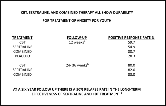
At 6-year follow-up, however, the data indicate that all treatments have equivalent relapse rates, of about 50 %, and presumably young adults with chronic and lifelong disorders (Ginsburg et al. 2014). That our treatments are not very effective is further suggested by studies of hospitalization and, more importantly, rehospitalization rates. Hospitalization rates for adolescents with psychiatric diagnoses have increased by 42 % over a 10-year period as reported by Bladder (2011). Further, rehospitalization rates, in Arnold et al. (2003) analyses, were in the 30–50 % range.
The problem, in short, is that we have been treating labels not the neurological condition.
The strategy of treating the condition and not the label is shown in the case example in Fig. 3.2. This client, a 51-year-old female, has a long history of “depression” that has not responded efficiently to any treatment, psychological or pharmaceutical. The problem is this client does not have depression markers; she is “depressed” because her life is in shambles resulting from severe anxiety and poor stress tolerance, identified in less than 15 min by a basic ClinicalQ assessment.
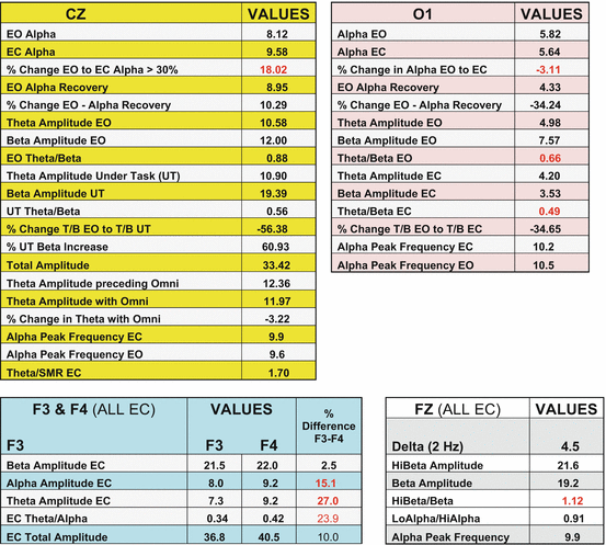
Fig. 3.2
Client with anxiety-based depression
Following the ClinicalQ probes, the client has trauma markers at both locations (Cz and O1), marked bilateral elevation of frontal Beta, marked deficiency in the Theta/Beta ratio at O1, poor sleep quality marker also at O1, marked elevation of 16 Hz through 40 Hz at Fz, emotional volatility marker at F4, and very good brain efficacy marker at Fz. All of these neurological conditions are a perfect storm for severe anxiety conditions. She is “depressed” because she cannot cope.
A female client in a hospital-based day program had a very similar ClinicalQ profile to the one shown in Fig. 3.2 above. She had major trauma markers at both locations, marked deficiency in the Theta/Beta ratio in the occipital region, marked elevation of frontal Alpha amplitude, as well as elevated 16–40 Hz amplitude at Fz.
This young woman was diagnosed with “pseudoseizure disorder,” often also called non-epileptic seizure disorder. Pseudoseizures are presumed to be sequelae of exposure to severe emotional trauma and may be related to the flashback process (see Swingle 1998a, b). In short, the pseudoseizure, which can be fainting, fugue-like state, tremors, and full-blown myoclonic like seizures, may occur to block the severe emotional distress associated with emotional flashback. The trigger for the emotion-blocking seizure may be physiological arousal as appears probable from the data shown in the following Fig. 3.3.
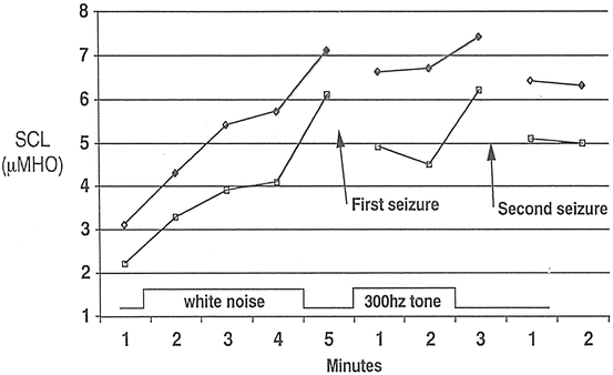
Fig. 3.3
Electrodermal conductance during auditory stimulation of a client with pseudoseizure disorder
During treatment sessions, seizures were common occurrences. This client had significant multiple medications, many to be used Prn, which was a major complicating factor in her treatment. During neurological/physiological and/or psychological/behavioral treatment sessions, this client’s physiological signs were monitored including electrodermal conductance, electromyographic amplitude (frontalis placement), peripheral blood flow (finger placements), heart rate, and blood oxygen saturation.
As shown in Fig. 3.3, seizure activity occurred when the electrodermal conductance rose above 6 micromhos bilaterally. The figure shows provoked activity associated with contingent sound (client’s tolerance of feedback and conditioned stimuli sounds to be used in neurotherapy was being evaluated), but similar physiological patterns were found in therapy sessions when strong emotion was elicited. Many readers will be familiar with the use of electrodermal (GSR) monitors during psychotherapy sessions to identify emergence of emotional content that may be important.
Depression
A common complaint of clients we all treat is “I’m depressed.” The client has a huge array of options for receiving treatment for this amorphous condition including prescription medications, supplements, exercise, endless psychotherapies, R & R, and of course an array of neurotherapies. In the latter case, we have normative database guided; neurofeedback, the z-score zapping paradigms (brain site-specific frequency amplitude departures penetrating z threshold evoke an infinitesimal amp/gauss zap), and z-score neurofeedback; sLORETA; canned feedback protocols based on defined condition (i.e., “depression”); and franchises with proprietary symptom checklist-driven canned protocol systems.
ClinicalQ-based treatment is different. The cases, described below, exemplify how treatment is guided by bottom-up assessment and verification. Neurotherapeutic protocols are then precisely targeted at these verified neurological inefficiencies. The ClinicalQs for the following cases are presented in summary form rather than the full output, as shown above. In addition, only data relevant to the present discussion are included in the summary.
The fundamental neurological condition one finds in depression is an imbalance in the frontal cortex with the right (F4) being more active as compared with the left (F3). The data presented in the previous chapter indicate that negative emotional states including feelings of depression, worthlessness, negative emotional life, nothing to look forward to in life, and fearfulness are all related to EEG data indicating that the right frontal cortex is more active than the left. There is substantial evidence for this relationship. An excellent review of the literature supporting the relationship between depression and elevated activity of the right relative to the left hemisphere (Hecht 2010) also reviews the evidence for comorbid conditions related to this imbalance as summarized in the abstract:
Depression is associated with an inter-hemispheric imbalance; a hyperactive right-hemisphere (RH) and a relatively hypoactive left-hemisphere (LH)… There is evidence that the RH is selectively involved in processing negative emotions, pessimistic thoughts and unconstructive thinking styles—all which comprise the cognitive phenomenology of depression and in turn contribute to the elevated anxiety, stress and pain associated with the illness. Additionally, the RH mediates vigilance and arousal which may explain the sleep disturbances often reported in depression. The RH had also been linked with self-reflection, accounting for the tendency of depressed individuals to withdraw from their external environments and focus attention inward. Physiologically, RH activation is associated with hypercortisolemia, which contributes to the deterioration of the immune system functioning and puts depressed patients at a greater risk of developing other illnesses, accounting for depression’s high comorbidity with other diseases. Conversely, the LH is specifically involved in processing pleasurable experiences, and its relative attenuation is in line with the symptoms of anhedonia that characterize depression. The LH is also relatively more involved in decision-making processes, accounting for the indecisiveness that is often accompanied with depression (Hecht 2010).
The imbalance between the right and the left frontal regions can result from several neurological conditions as measured with the EEG. The Davidson (1992) pattern, identified years ago, is when Alpha has greater amplitude in the left relative to the right. However, there are many other conditions that result in this imbalance. For example, the client shown in Fig. 3.4 is what we might call “garden variety” depression. This client has an imbalance where Beta is greater in the right relative to the left. Clinically this appears to be the “genetic” predisposition for depression although it is found in client’s having recently experienced a loss. Figure 3.5 shows the Davidson depression marker of elevated Alpha in the left relative to the right frontal cortex. The client shown in Fig. 3.6 is similar in that Theta is greater in the left relative to the right resulting in the right being more active than the left. Clinically the two patterns just described (low-frequency amplitude greater in the left) are very frequently associated with reactive depression (exogenous). Finally in Fig. 3.7, we see a pattern often found with a person with the predisposition to depression who has experienced a severe emotional stressor that has triggered the predisposition.
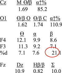
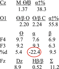
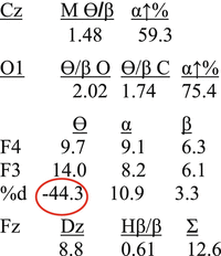
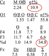
Fig. 3.4
“Genetic” depression
Fig. 3.5
Reactive depression (Alpha)
Fig. 3.6
Reactive depression (Theta)
Fig. 3.7
Trauma-triggered depression
Emotional trauma, exposure to a severe emotional stressor or an accumulation of emotional stressors, is associated with a blunting of the Alpha response at locations Cz and O1. We understand that this marker is associated with incompletely processed emotional sequelae of the emotional event(s). Exposure to emotionally negative images (corpses) has been shown to temporarily blunt the Alpha response and fortuitous exposure to severe emotional stress with clinical clients likewise revealed Alpha blunting. Alpha blunting is seen as restricted elevation of Alpha amplitude when clients close their eyes (Swingle 2013) (see the parameters for this response in the Appendix to this chapter). The Alpha response is completely ignored in the normative databases.
Occasionally one sees clients who report that they are depressed but there are no depression markers in the ClinicalQ. There are many profiles that are found but two are relatively common. The profile shown in Fig. 3.8 shows no depression markers but both trauma markers. There are other details of clinical relevance in this profile, but the critical point for this discussion is that unprocessed trauma can be manifested as reports of “depression.” The lack of the reactive depression markers (e.g., Davidson 1992) may indicate that the client is in the numb phase of posttraumatic exposure. However, although of interest to speculate on these matters, clinically one proceeds to release the Alpha and then utilize whatever therapy the clinician judges relevant to resolve the condition. It is with these trauma clients that the one-size-fits-all franchisers are the most destructive. Often one will hear comments about how to quiet an emotionally abreacted client who has been subjected to one of the canned protocols, exactly the opposite of good clinical practice.
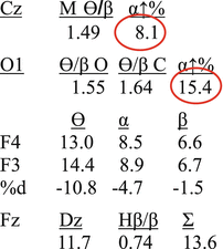
Fig. 3.8
Trauma-based depression
The profile shown in Fig. 3.9 is also quite common. These are clients in severe states of anxiety who feel hopeless, frightened, and out of control. They report being “depressed” because their lives are in shambles, or they feel they are going to decompensate, or they feel just plain helpless. Treating these conditions with antidepressants is a formula for creating a lifelong problem. The ClinicalQ identifies the areas for neurotherapeutic treatment quite precisely. Again, there are several other aspects to this EEG profile of clinical relevance such as markers for cognitive perseveration, but for the purposes of the present discussion, it is the two markers of deficient Theta/Beta ratio at the occipital location and elevated left frontal Beta that identify the anxiety state.
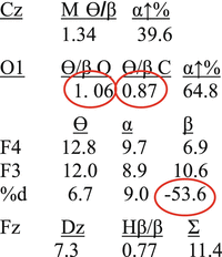
Fig. 3.9
Anxiety-based depression
As noted in earlier sections, clients who rate themselves high on “depression” have, on average, 10 % lower amplitude Theta/Beta ratio at location O1 relative to those clients who rate themselves not depressed (t = 2.20, p < 0.03, df = 367). In addition, the correlation between the ratings of “I feel depressed” and “I am very anxious” is 0.51 (p < 0.0001, df = 912), indicating that many clients who report depression may well be experiencing the sequelae of severe anxiety without any of the neurological markers for depression. In our experience at the Swingle Clinic, these are the clients who frequently report histories of many years of “depression,” many years of therapy, and many different pharmaceutical cocktails. Obviously, neither the therapies nor the pharmaceuticals were satisfactory; otherwise they would not be seeking treatment. The problem, again, is that the treatment of these clients was unsuccessful because the label not the neurological condition was being treated.
Another common profile associated with client complaint of “depression” is associated with misdiagnosed and mistreated high frontal Alpha form of ADHD (see Fig. 3.10). This case will be reviewed more thoroughly in the next section. The depression associated with this condition is reactive because the client feels out of control, emotionally dysregulated, and disheartened. Frequently and unsuccessfully treated with a variety of psychotherapies and/or pharmaceutical cocktails usually including both antidepressants and anxiolytics (and often with atypical antipsychotics as well), these clients often have treatment histories that span decades. As the above ClinicalQ profile indicates, this client’s major neurological condition is marked excessive Alpha amplitude over the frontal cortex. It is ADHD, in other words, and this client would have been spared decades of angst if the correct condition, not the label, had been treated.
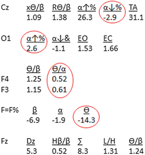
Fig. 3.10
High frontal alpha-based depression
The treatment of children with early signs of depression can be particularly challenging. In the first instance, it is very likely that the indicators will be missed or interpreted as some other condition such as anxiety, ADHD, and even mild autism. Depression in children is often not associated with outward indicators of sadness that we associate with depression. These children often seem to be lacking energy, interest, and motivation. They do not appear interested in the activities in which they are engaged. However, these characteristics can also be related to sleep problems or anxiety. As detailed in the last chapter, children with deficient Theta/Beta ratios in the occipital region of the brain are more likely to be rated, by a parent, as having a sleep problem and lower in self-esteem. They are also rated higher on “unhappy,” “poor self-esteem,” “indifferent,” “frequently ill,” and “easily frightened.”
Many of the above characteristics can also be considered symptoms of depression. In a study of preschoolers (Luby et al. 2014), children diagnosed with depression were 2.5 times more likely to be diagnosed with major depression disorder at school age. However, the authors note that preschool depression does not meet the criteria for major depression disorder. They suggest that onset of preschool depression may be more common than is clinically recognized. These children show lack of joyfulness during normal play rather than frank sadness, and the symptoms appear to be intermittent. This is related to the finding from the ClinicalQ database indicating that children with the low Theta/Beta ratio at the occipital site show more “indifference” which seems consistent with the lack of joyfulness reported by Luby et al. (2014).
The above data make it quite clear that one should treat the putative neurological condition and not the symptoms or diagnostic label. Anxious and sleep-disturbed children may show many of the signs of depression, as do adults, and be inappropriately treated for depression. Following the neurological data from the ClinicalQ directs treatment to the functional brain inefficiency, regardless of label, that can be treated to improve the client’s condition.
In addition to the relationship between reported anxiety and reported depression, anxious clients also rate themselves higher on fatigue (r = 0.39), perseverative thoughts (r = 0.47), negative view of their future (r = 0.40), irritability (r = 0.39), problems with concentration (r = 0.33), and feelings of worthlessness (r = 0.45). These associations are quite consistent with the correlations with self-rated depression: fatigue (r = 0.52), perseverative thoughts (r = 0.42), negative view of the future (r = 0.56), irritability (r = 0.38), and problems with concentration (r = 0.37). In addition, self-rated depressed clients are more likely to rate themselves as feeling physically unwell (r = 0.36). All of the correlation coefficients are statistically significant (p < 0.0001, N = 914).
Recall the data on manifestation of neurological predispositions such as in the study of monozygotic twins, one of whom had manifested schizophrenia, whereas about 50 % of the twin did not. Depression markers are indicators of predisposition. They are markers for neurological conditions that can manifest to clinical level depression. But many will not. This is one of the principal reasons why normative databases are notably inaccurate at detection of clinical conditions, whereas clinical databases are considerably more useful for the clinician. However, even in clinical populations, some clients presenting conditions other than depression might also have markers for unmanifested conditions. Clients with no markers for depression distribute themselves about equally among low, mid, and high self-ratings on the depression items on the intake questionnaire. About 32 % rate themselves as high, 34 % as mid-range, and 33 % as low on “I feel depressed.” Alternatively, 26.3 % of clients with at least one depression marker rate themselves as without depressed mood states. These are the clients with neurological markers that, like the unmanifested genetically predisposed schizophrenic, have not been exposed to conditions that result in symptom manifestation.
Bipolar Disorders
Many clients come with diagnosis of “bipolar” disorder. When questioned, many state that they have a history of depression but no recall of any “manic” phases. Others feel that the hyper states have been the primary cause of their difficulties in all aspects of their life. In keeping with the therapeutic dictum of “treat the condition not the label,” the ClinicalQ is used to identify neurological conditions that may predispose a client to depressed, agitated, and emotionally dysregulated states. Clients may report symptoms of depression resulting from feeling hopeless, powerless, or stupid because of irrational behavior and emotional states associated with the manic phases in some forms of bipolar disorder. On the other hand, bipolar diagnoses have been attached to conditions that are actually high frontal Alpha forms of ADHD.
From the clinical database, 23 clients came for treatment with a diagnosis of “bipolar disorder” from an independent psychiatrist. Of this group of clients: 91.3 % had one or both of the trauma markers (blunting of the Alpha response at locations Cz and O1) as compared to 39.9 % in the general clinical group (z = 6.77, p < 0.001, N = 891); 87.0 % had at least one of the three emotional volatility or dysregulation markers (elevated Alpha amplitude at F3 and F4, elevated right frontal Theta amplitude at F4, or elevated right frontal Alpha amplitude) as compared to 68.3 % in the general clinical group (z = 2.03, p < 0.05, N = 891); 82.6 % had the poor stress tolerance marker (deficient Theta/Beta ratio at O1) as compared to 71.4 % in the general clinical group (z = 1.18, ns); 58.0 % had at least one of the four depression markers (elevated F4 Beta amplitude, elevated F3 Theta and/or Alpha amplitude, elevated Theta/Beta ratio at F3) as compared to 37.0 % in the general clinical group (z = 2.01, p < 0.05, N = 891). It is interesting to note that these data are not inconsistent with common pharmaceutical treatments for these conditions that often include mood stabilizers, antidepressants, and anxiolytics. It is also interesting to note that the principal discriminators for the bipolar group were the trauma markers and the markers for emotional volatility and dysregulation. About 90 % of the bipolar clients had trauma markers, and about the same percentage had an emotional dysregulation marker. Further 100 % of this group had at least one of these markers.
About half of the bipolar clients had at least one marker for depression as compared with about a third of the general clinical group. It is apparent then that the major discriminative neurological features of the bipolar group are emotional volatility markers and indicators of exposure, past and/or present, to severe emotional stress.
There also appears to be an elevated connectivity among brain sites. Correlations between frontal sites F3 and F4 for Theta, Alpha, and Beta amplitude, the ratios of Theta/Beta and Theta/Alpha, and correlations between these frontal sites and the SMR at location Cz were all substantially higher in the bipolar group as compared with the general database. Of the 17 correlations, 16 were greater (direction ignored) for the bipolar group (binomial probability, p < 0.001). The difference between the average correlations for the general group (r = 0.29, SD = 0.16) and for the bipolar group (r = 0.79, SD = 0.21) is statistically significant (t = 6.26, p < 0.001, df = 20). The coefficients are also significantly different (recognizing that these are averages, direction ignored) based on sample size of 16 (z = 3.66, p < 0.001).
There was a nonsignificant trend for a greater proportion of the bipolar group to have markers for poor stress tolerance which was high for both groups. In this regard, it is interesting to note that adolescents diagnosed with a bipolar disorder have a higher probability of developing a substance use disorder (SUD), most frequently cannabis. In a sample of bipolar adolescents hospitalized with mania, 48 % had or developed an SUD (Stephens et al. 2014). This high rate of SUD is consistent with the high rate of deficient Theta/Beta ratios found in our sample of clients diagnosed with a bipolar disorder.
It is also apparent that the neurology is not the only area of concern in dealing with these clients. As is discussed throughout this book, the efficient processing of the sequelae of exposure to severe emotional stress (trauma) is central to the effective treatment of these clients. Similarly, some focus on developing self-regulation procedures for dealing with emotional dysregulation is central. This is particularly problematic because these dysregulated behaviors are likely to developed stability because of conditioning. The common example is the emotionally volatile individual who’s unpredictable and explosive emotional tirades have been reinforced by being instrumental to the control of others.
Two clients, both of whom were diagnosed as bipolar, showing different ClinicalQ patterns are shown in the following two figures. Figure 3.11 shows the trauma marker at Cz, the deficient Theta/Beta ratios at O1 (with the sleep quality problem marker), the frontal depression marker (elevated right frontal Beta), and two emotional/mood volatility markers (elevated right frontal Theta and Alpha amplitudes).
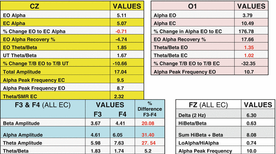
Fig. 3.11
Female, 37 years old, client diagnosed with bipolar disorder
The second case, shown in Fig. 3.12, is the rare bipolar client that does not show trauma markers. This client rated herself at maximum scale levels for depression, anxiety, fatigue, perseverative thoughts, feelings of worthlessness, and problems with concentration and sleep. This client has the high frontal (F3 and F4) Alpha amplitude that is associated with both cognitive and emotional dysregulation.
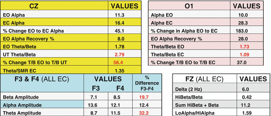
Fig. 3.12
ClinicalQ summary of 51-year-old male diagnosed with bipolar disorder
These individuals have problems with planning, organizing, and following through and problems with concentration and focus. They also often have major problems with emotional dysregulation with irritability, mood swings, and volatility. The profile also shows marker for sleep disturbance (deficient eyes closed Theta/Beta ratio at O1), depression (elevated Beta amplitude at location F4), and an additional marker for mood volatility (elevated Theta amplitude at F4). This client also shows the marker for another form of ADHD and a particularly troublesome one. Note that the Theta/Beta ratio is in an acceptable range under at-rest conditions but jumps substantially when the client was under cognitive challenge (reading out loud). This is very problematic, for the brain, although seemingly within normal limits, produces substantial Theta amplitude when the client is attempting to focus. This is often associated with subjective feelings that conditions get worse the harder the person tries to concentrate (very discouraging for a school-age child).
Obviously the treatment for these two clients will be quite different. The first client is likely to have emotional abreactions when the Alpha response is restored as the emotional properties of the memories of the traumatic event start to emerge. Both clients have markers for depression and sleep problems, but the second client also has two markers for different forms of ADHD, one of which, as stated above, is also related to emotional dysregulation. One often sees clients with major depression complaints who show these ADHD markers but often no depression markers. This is usually associated with problems with life discouragements including academic, career, and interpersonal relationships. The “depression” is associated with the feelings of failure, despair, helplessness, hopelessness, and confusion. These clients often do not respond well to antidepressant medications and often have long histories of psychotherapy and psychopharmacology. The lack of meaningful progress with these therapies further contributes to feelings of despair and confusion.
The Attention Deficit Hyperactivity Disorders
In this section, we cover the disorders that are, indeed, disorders of attention. In this category we examine the neurological conditions that directly affect attention and hyperactivity. These are situations in which the symptoms are not primarily the result of some other condition such as depression. Much has been written about various forms of ADHD, and we find that researchers and clinicians differ in the number they suggest. Most of these variations in forms of ADHD are really ADHD plus various comorbid conditions such as sleep disturbance, depression, oppositional behavior, anxiety, and the like. As the reader will find, the labels and “types” of ADHD are largely irrelevant. From the perspective of “bottom-up,” we look at the neurology of the child and correct relevant anomalies in functioning.
In latter chapters, the conditions that interfere with a child’s ability to be attentive in school but are not neurological disorders of attention are reviewed. These will include conditions of poor stress tolerance, predisposition to depression, oppositional and defiance disorders, and frightened and traumatized children.
One of the problems we have with the statistics associated with consequences of untreated ADHD is that the methods for diagnosing these conditions are so flawed. As pointed out in an earlier chapter, the top-down method for diagnosis of attention deficiencies is simply inappropriate. However, with this in mind, let us remember some of the sequelae of untreated attention problems.
There is disagreement among researchers about the risk factors associated with untreated ADHD. Part of this problem, of course, is the fact that ADHD is a “wastebasket diagnosis.” However, most of the disagreement is not about the fact that untreated ADHD leads to life complications; rather, that this disagreement centers on the extent of the risk. And as we shall review at various times throughout this book, different forms of ADHD pose different risks for individuals who remain untreated into adulthood. Other factors, of course, are important, including any psychological comorbidity one might have such as depression and anxiety, as well as gender and culture, to name but a few.
Although there are some inconsistencies in the data, nonetheless a relationship between untreated ADHD and criminality seems clear. Data collected on incarcerated males indicate much higher proportions of those with ADHD-like behaviors than in the general population. Interestingly, these individuals are not only more likely to be charged with a crime and arrested, but they are also more likely to be indicted and far more likely to be imprisoned. Perhaps this simply indicates again that they lack the capability for responsible planning, organizing, sequencing, and monitoring their personal situation once they have been charged with a crime.
A study, by Dr. Ginsberg and colleagues, of long-term male inmates in a Swedish prison found that 40 % had ADHD and less than 7 % had ever been diagnosed with this condition. Another study, by Drs. Fletcher and Wolfe of Yale University, of 13,000 adolescents over a long time period found that those with ADHD were twice as likely to commit a robbery and 50 % more likely to have sold drugs, as compared to their peers.
So, it seems clear that undiagnosed and untreated ADHD is a very substantial risk factor for criminal, and otherwise irresponsible, behavior. The challenge is to make sure that we treat the behaviors that are causing the trouble and not be corralled by the label we put on a specified grouping of these troublesome behaviors. Doing the latter markedly reduces our success rate for helping children overcome these hurdles to successful and fulfilling lives.
The data shown in Table 3.1 come from various studies of adults with untreated ADHD (Biederman et al. 2006; Kessler et al. 2006; Rösler et al. 2004; Young et al. 2002). And although one can quibble about the magnitude of some of these findings, nonetheless the data are striking. The following table gives some statistics from various studies of the consequences of untreated ADHD. This is a table of adult populations of individuals whose ADHD was either not diagnosed properly and/or did not receive proper and efficient treatment for the condition.
Table 3.1
Sample studies of adult populations with untreated ADHD conditions
ADHD-associated risks |
|---|
Social/emotional/addiction problems
Related posts:Stay updated, free articles. Join our Telegram channel
Full access? Get Clinical Tree
 Get Clinical Tree app for offline access

|


