Chapter 20 Musculoskeletal ultrasound imaging
INTRODUCTION
Ultrasound imaging enables the clinician to look directly at the soft tissues of the body. The dynamic, real-time, moving image that is produced is ideal for evaluating the structure and behaviour of muscles, tendons and other soft tissues. Not only can ultrasound be used to evaluate pathological changes, it is also possible to analyse muscle contractions, the movement of tendons and joints, and their effect on surrounding structures.
The relatively low cost and safety (owing to the absence of ionizing radiation) of ultrasound scanners make them a practical tool in the clinical setting and, unsurprisingly, they are no longer the preserve of radiology and maternity departments. For many clinicians, such as cardiologists, anaesthetists and surgeons, using ultrasound is now seen as a natural extension of their clinical examination in much the same way as the stethoscope.
The use of ultrasound imaging in physiotherapy is still in its infancy but has already developed applications beyond those traditionally performed in radiology. Monitoring response to treatment, along with evaluating function and the dynamic interaction between structures, is often of more relevance to the therapist. This chapter introduces the reader to ultrasound imaging and highlights those applications that might be relevant to physiotherapy practice.
HISTORY
The use of ultrasound to image the human body began shortly after the Second World War, using technology borrowed from military and metallurgy applications. Early studies required the subject to be immersed in a water bath, and produced a single, static, cross-sectional image.
By the early 1980s, a moving image could be produced. This was acquired by applying a small probe to the skin surface, which allowed quick and straightforward examinations of many of the organs of the body. At around this time CT and MRI also became available as alternative methods of producing cross-sectional images. Each modality was found to have its own strengths and weaknesses and, despite rapid developments over the past 20 years, they remain complementary, rather than rival approaches to imaging.
Musculoskeletal ultrasound applications began to emerge in the mid-1980s (Crass et al 1984) as the resolution of scanners improved. Physiotherapy researchers found ultrasound to be ideal for the direct visualization and evaluation of both muscle size and activity (Stokes&Young 1986). By the late 1990s, the development of high-frequency probes, with resolution in some respects superior to MRI, had led to renewed interest in musculoskeletal ultrasound. This continues today as falling costs and increased portability have made high-resolution ultrasound imaging feasible in the clinic and on the ward.
PRINCIPLES
GREY SCALE
When a sound wave passes through the body, a small percentage of the wave’s energy (typically less than 1%) is reflected at each boundary that is encountered. The ultrasound image is acquired using a probe that generates a series of short, tightly focused pulses. These pass through the body and the sound waves reflected back to the probe by successive boundaries are recorded. Each pulse produces a single line of data, and a single image may be made up over a hundred such lines.
With each image taking as little as 1/100th of a second to construct, it can be updated continuously, giving a real time cinematic view of the body that allows even fast-moving structures such as the heart, active muscles, or even small children to be imaged.
SCANNERS
Scanners are typically made up of one or more hand-held probes attached via flexible cables to a central unit, which processes and displays the image (Fig. 20.1). The probes operate at one or more fundamental frequencies, which determine the maximum depth and resolution of the images they generate. At higher frequencies, the shorter wavelengths mean that shorter pulses can be produced, giving better resolution. At lower frequencies, the resolution is reduced but the ultrasound is absorbed less readily as it passes through the body, enabling deeper structures to be examined.
The most commonly used probes are the linear and curvilinear probes (Fig. 20.1B). Linear probes are normally designed to operate at frequencies of 7.5 MHz and above; these produce high-resolution, rectangular images to a depth of around 5 cm. Curvilinear probes generate a fan-shaped image with a wider field of view than a linear probe. They operate at lower frequencies, typically between 2 and 5 MHz, and can produce images to a depth of up to 15 cm but at the cost of lower resolution.
A detailed discussion of resolution and image generation lies beyond the scope of this chapter and the reader will find more comprehensive descriptions in dedicated ultrasound texts, in particular Hedrick et al (2005).
DOPPLER
Doppler is used to detect the flow of blood within the body by measuring the change in frequency of a sound wave that occurs when it is reflected by a moving structure. The two common display modes are colour and pulsed Doppler.
Colour Doppler
This samples the flow velocities over a wider area of the image and displays the presence of flow by adding colour to the greyscale image (Fig. 20.2A). Blue and red are typically used, although these represent flow either away or towards the ultrasound probe rather than venous and arterial flow.
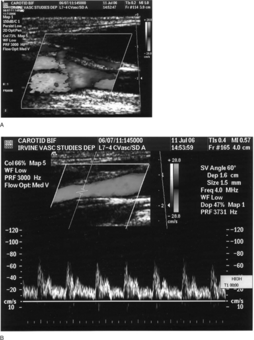
APPEARANCES: FEATURES OF AN ULTRASOUND IMAGE
Presentation
The orientation of the screen is not fixed but for most applications the top of the image corresponds to the surface in contact with the ultrasound probe. For ultrasound applications such as abdominal scanning, the convention used in CT and MRI is generally adhered to, with the left side of a transverse section image corresponding to the right side of the patient. This is done so that the image corresponds to what the clinician sees when facing the patient, looking up from the feet. By convention, the left side of a longitudinal or coronal ultrasound image represents the cephalic direction, which corresponds with what would be seen by an observer looking from the right side of the patient. For musculoskeletal scanning these conventions are not always appropriate as the position of the patient and operator are not standardized, though ambiguous images should be adequately labelled.
The depth of the image is displayed as a scale of distance from the probe, running down the side of the picture. The maximum depth that can be displayed is determined primarily by the frequency the probe operates at, but is also dependent on the power output, and by how strongly the sound is absorbed (attenuated) by the intervening tissue.
Typical appearance of normal tissue
The normal appearance of structures varies from person to person. The frequency used and the quality of the scanner also determines the amount of detail in the image. The most significant factors affecting the image, however, are the amount and nature of tissue that the sound has to pass through to reach the structure of interest.
Figure 20.3 demonstrates some typical appearances of soft tissues:

Stay updated, free articles. Join our Telegram channel

Full access? Get Clinical Tree

