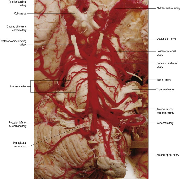
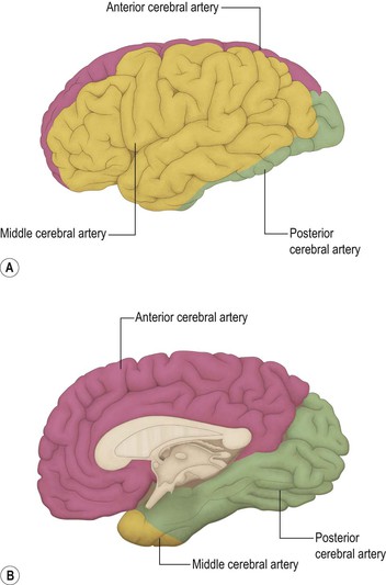
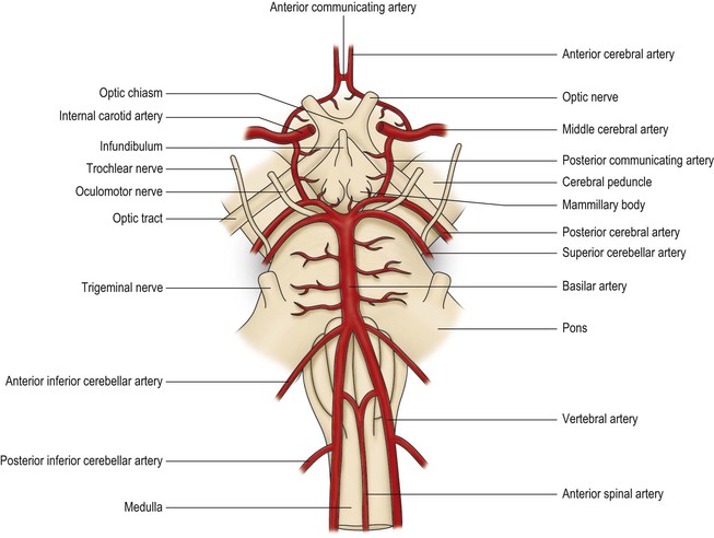
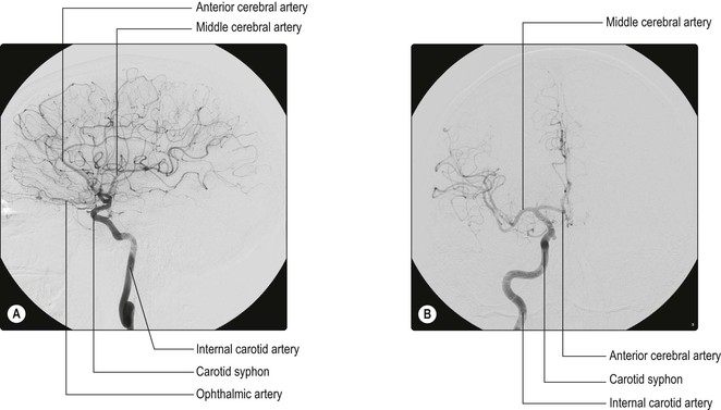
Three longitudinal arterial vessels run the length of the spinal cord (Fig. 7.1). These are the single anterior spinal artery and the paired posterior spinal arteries. The anterior spinal artery arises in a Y-shaped configuration from the two vertebral arteries at the level of the medulla oblongata (Fig. 7.2) and descends along the ventral surface of the cord in the midline. The posterior spinal arteries arise from either the vertebral arteries or the posterior inferior cerebellar arteries and run caudally on the posterolateral surface of the cord. The anterior and posterior spinal arteries alone are insufficient to supply the cord below cervical levels and, therefore, they receive serial reinforcement by anastomosis with radicular arteries derived from segmental vessels, including the ascending cervical, intercostal and lumbar arteries. Radicular arteries pass through the intervertebral foramina and divide into anterior and posterior branches, which run with the dorsal and ventral spinal nerve roots, respectively. One particularly large radicular artery (the great radicular artery, or artery of Adamkiewicz) may arise from a lateral intercostal or lumbar artery at any level between T8 and L3. The blood supply of the spinal cord is most vulnerable in the thoracic region and in the anterior portion of the cord. Occlusion of the anterior spinal artery leads to an acute thoracic cord syndrome with paraplegia and incontinence. The spinothalamic modalities of pain and temperature are preferentially lost, whereas the proprioceptive functions of the dorsal columns are relatively preserved. The venous drainage of the cord follows a basically similar pattern to the arterial supply (Fig. 7.1). Six longitudinal interconnecting venous channels exist. These consist primarily of anterior and posterior spinal veins, which run in the midline. More irregular, sometimes incomplete, bilaterally paired anterolateral and posterolateral veins are situated near the lines of attachment of the ventral and dorsal nerve roots, respectively. All of these vessels drain via anterior and posterior radicular veins into the internal vertebral venous plexus (epidural venous plexus), which is situated between the dura mater and the vertebral periosteum. The internal venous plexus communicates with an external vertebral venous plexus and thence with the ascending lumbar veins, the azygos and hemiazygos veins. The brain is supplied with blood by two pairs of vessels, the internal carotid arteries and the vertebral arteries (Figs 7.2, 7.3 and see Fig. 7.7). The internal carotid artery arises from the common carotid artery and enters the middle fossa of the cranial cavity through the carotid canal. Its course then follows a series of characteristic bends, known as the carotid syphon (Fig. 7.4), after which it passes forwards through the cavernous sinus and then upwards on the medial aspect of the anterior clinoid process, reaching the surface of the brain lateral to the optic chiasm. Along its course, the internal carotid artery gives rise to a number of pre-terminal branches. ▪ Hypophyseal arteries arise from the intra-cavernous section of the internal carotid to supply the neurohypophysis. They also form the pituitary portal system of vessels by which releasing factors are carried from the hypothalamus to the adenohypophysis (Ch. 16). Lateral to the optic chiasm, the internal carotid artery divides into its two terminal branches, the anterior and middle cerebral arteries. The anterior cerebral artery courses medially above the optic nerve and then passes into the great longitudinal fissure, between the frontal lobes of the cerebral hemispheres. As it does so, it is joined to the corresponding vessel of the opposite side by the short anterior communicating artery. Within the great longitudinal fissure, the anterior cerebral artery follows the dorsal curvature of the corpus callosum (Fig. 13.23), branches ramifying over the medial surface of the frontal and parietal lobes, which it supplies (Fig. 7.5). The territory supplied by the anterior cerebral artery, therefore, includes the motor and sensory cortices for the lower limb. Fine terminal branches also extend out of the great longitudinal fissure to supply a narrow lateral band of frontal and parietal cortices.
Vasculature of the central nervous system
Vasculature of the spinal cord
Arterial supply of the spinal cord
 Disorders of the blood supply of the spinal cord
Venous drainage of the spinal cord
Vasculature of the brain
Arterial supply of the brain
< div class='tao-gold-member'>
Related posts:
![]()
Stay updated, free articles. Join our Telegram channel

Full access? Get Clinical Tree


Vasculature of the central nervous system
Chapter 7

Only gold members can continue reading. Log In or Register to continue


