Ventral and Lateral Thoracic and Lumbar Fixation Techniques

Chapter 146 Ventral and Lateral Thoracic and Lumbar Fixation Techniques


Surgery on the ventral thoracic and lumbar spine began nearly 100 years ago. Ventral approaches for decompression of spinal pathology were first attempted in the early 1900s. Pioneers such as Royle1 excised hemivertebrae for the treatment of scoliosis. Ito2 as well as Hodgson and Stock3 refined the ventral (transperitoneal) approach to the thoracolumbar spine for the treatment of Pott disease. These early efforts to decompress ventral spinal pathology were frequently complicated by postoperative mechanical instability and progressive deformity.


The first reports of ventral instrumentation of the spine were from Humphries,4 who developed ventral interbody fusion with ventral plates and unicortical screws. These devices provided little biomechanical advantage. Most of these cases were transperitoneal approaches for debridement and stabilization in patients with Pott disease. The transperitoneal approach was eventually replaced by the retroperitoneal or extracavitary approaches for lesions of the lower thoracic and lumbar spine.


Throughout the 1970s, the preferred treatment for traumatic injuries was dorsal fusion and instrumentation, combined with ventral decompression and fusion. The Dwyer57 and Zielke8 devices were developed as ventrolateral implants that could augment or replace dorsal instrumentation. These consisted of screws that traversed the vertebral body that were interconnected with cable (Dwyer) or threaded rods (Zielke) that could be tightened in tension. These devices had limited ability to fixate two-column traumatic injuries. The Dunn device (developed in the late 1970s) represented a more rigid instrument for use in burst fractures. This double-screw, double-threaded rod device provided excellent strength but was removed from the market in 1986 after reports of great vessel erosion and rupture.9


It was not until the 1990s that numerous plate-screw and screw-rod systems were developed. Among the first were the Kaneda, Kostuik-Harrington, I-plate, and University plate systems. Later products included the anterior locking plate, the Z-plate, the Texas Scottish Rite Hospital system, the M-8 dual-rod system, the Expedium Anterior system, and others. Newer systems were developed for lower-profile, rigid ventral fixation of the spine. These systems have the added advantage of easier means of distraction and compression at the moment of plate or rod fixation. Furthermore, shorter segment fusions became more feasible. Some of the early generation ventral plating systems did not provide for a fully rigid screw-plate interface. Bicortical screw purchase was important but did not eliminate the possibility of screw toggle and possible mechanical failure in the early systems. However, most systems on the market by 2003 provided for a rigid screw-plate or screw-rod interface. To analyze the various systems critically, an appreciation of the biomechanics of the thoracolumbar spine is important. The reader is directed to other chapters outlining more detailed biomechanical information. It is also essential to understand some of the general indications for ventral instrumentation and fusion.


Biomechanical Considerations


A complex discussion of ventral thoracolumbar instrumentation is beyond the scope of this chapter; however, recognition of several basic concepts is essential to avoid complication. The anterior and middle columns provide the most resistance to axial loads. The ventral approach to spinal pathology has the distinct advantage of allowing for reconstruction of the vertebral body and intervertebral space with autologous or allogeneic bone or synthetic materials (e.g., methylmethacrylate, ceramic). The dorsal tension band—normally provided by the interspinous ligaments, ligamentum flavum, paraspinal musculature, and facet joints—must not be severely damaged if a ventral construct alone is to provide stability, because these devices cannot effectively withstand tremendous flexion forces. Complications arise when a posterior column injury has gone unrecognized or when too much is expected of a ventral construct. Plate and screw constructs can provide resistance against axial load, distraction, and extension. These implants have a higher success rate when axial loads are shared by a sturdy bone graft and the implant.


Certain biomechanical characteristics of implant systems are important to understand. Rigid implants (e.g., Z-plate, Kaneda systems, Expedium Anterior system, and M-8 dual-rod system) theoretically allow for greater immobilization of the spine. If the implant bears most of the stress, there is a risk of implant breakage or failure. Some plate systems have set screw holes rather than slots, thereby creating a static (nondynamic) condition beginning at the time of plate fixation. Also, stress shielding provided by the rigid fixation may prevent the beneficial compressive forces from enhancing bone fusion (Wolff’s law). Because bone is a biologic, deformable material, repeated stress loading may cause bony erosion and failure at the metallic implant-bone interface.


Indications for Ventrolateral Instrumentation


A variety of infectious, neoplastic, congenital, and traumatic pathologic conditions are suitable for ventral thoracic or lumbar surgery. An initial step in complication avoidance is to determine whether a ventral approach truly provides the safest and most efficacious means of decompression of the neural structures, reconstruction of the anterior and middle column, application of corrective forces for realignment, and placement of an appropriate graft or spacer-implant construct. Conditions in which dorsal neural compression or posterior column bony/ligamentous damage are the predominant findings are best treated by a dorsal approach. Similarly, lesions with three-column pathology may possibly require circumferential treatment.


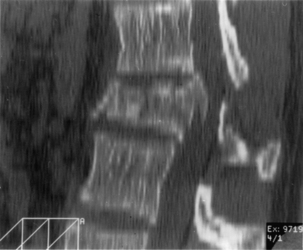
Anterior and middle column trauma (with preservation of the dorsal elements) may be treated adequately with a ventral approach (Fig. 146-1). Failure to recognize significant posterior column injury may result in delayed kyphotic deformity and neurologic deterioration. There are few clinical outcome data to encourage ventral decompression of trauma patients with complete neurologic loss below the level of the lesion. Ventral approaches, however, may be useful in paraplegic patients with a severe kyphotic deformity. Anterior reconstruction may provide better sagittal balance that could be important for long-term pulmonary function, independent transferring, and upper extremity function.


Patients with incomplete spinal cord injury, severe vertebral body collapse (≥40%) and kyphosis, and/or significant spinal canal compromise should be considered for ventral decompression and reconstruction. Intact patients with a myelographic block or ventral compression of the spinal cord on MRI may be considered for ventral decompression and stabilization. If the posterior longitudinal ligament (PLL) is intact on MRI and there is 30% or less loss in height of the anterior and middle column, a dorsal approach with reduction by ligamentotaxis could be considered in the intact or incomplete patient with a burst fracture.


Infection is another indication for ventral decompression, reconstruction, and instrumentation. The primary indication would be severe deformity of the spine, because most spinal infections can be treated without ventral instrumentation. Early ventral approaches for the treatment of Pott disease have been modified and are still very useful in debridement and stabilization of pyogenic, mycobacterial, or fungal infections. Reconstruction with autologous or allogeneic bone is feasible if a complete debridement of all necrotic tissue is accomplished. The risk of persistent infection or implant failure with instrumentation of infected cases is low if a prolonged course of antibiotics is given to these patients.


Metastatic neoplasms commonly affect the vertebral body before the posterior elements and therefore can be palliated with vertebrectomy. The cell kinetics of any malignancy, if known before surgery or determined by frozen section, must be considered when deciding on the material for reconstruction of the axial spine. In cases of a rapidly dividing carcinoma, synthetic spacers such as vertical titanium cages, methylmethacrylate, polyether ether, or carbon fiber cages can provide immediate stability of the vertebral column in patients with a short life expectancy. In more indolent neoplasms, such as breast or prostate cancer, longer survival can be expected, and autogenous or allogeneic bone can be expected to incorporate, thereby avoiding complications such as pseudarthrosis or implant migration. Use of titanium and other nonferromagnetic implants allows for long-term follow-up with MRI.


Treatment of other conditions, such as congenital or developmental scoliosis, iatrogenic lumbar kyphosis (flat back syndrome), and degenerative lumbar scoliosis may involve a ventral approach. Ventral procedures in adult scoliosis with curves greater than 40 to 50 degrees are associated with a higher rate of fusion than dorsal constructs alone. Ventral fusion and instrumentation may also be useful in patients with deficient laminae, facet joints, or pars interarticulari or extremely severe scarring from prior dorsal surgery.


Inadequate radiographic studies before surgery can lead to intraoperative or postoperative complications. Plain radiographs are essential and should include flexion and extension views when there is any suspicion of mechanical instability. In addition, attention should be paid to the density of bone as well as the sagittal and coronal alignment. Patients with scoliosis should have complete 36-inch standing films to assess overall spinal balance. The value of sagittal reconstruction of CT images is often overlooked, particularly after myelographic dye injection. Axial CT may be preferable over MRI to determine the amount of bony spinal canal compromise in trauma. Sagittal MRI often provides excellent views of the PLL. If intact, one may consider use of ligamentotaxis to reduce a burst fracture fragment. MRI has the added advantage of showing signs of soft dorsal tissue injury and hematoma that would commonly go unrecognized with plain radiographs and CT scan alone. Although cost-effectiveness is a primary concern, any patient with complex spinal pathology (and for whom aggressive surgery is contemplated) may require both CT and MRI as part of the workup.


Ventral Surgical Techniques


Positioning


Complication avoidance in the operating room begins with the simplest of steps. In positioning all patients, foam rubber padding is placed over ankles and elbows. For the prone position, gel pads are placed over the supports of the Wilson frame or chest rolls. The knees are flexed 45 to 90 degrees. Electrocardiography electrodes must not be on areas of the chest or trunk that contact the frame or rolls to prevent pressure necrosis. In females, the breasts must be tucked medially between the supports. Pillows are placed under the feet to provide knee flexion and relaxation of the sciatic nerve. Foam rubber rings are commonly used by anesthesiologists to protect the face and eyes, but care must be taken not to place the patient’s neck in too much extension when in the prone position, particularly in one with diffuse spondylosis. It is necessary to double-check the eyes to ensure that there is no direct pressure on the globe. If the arms are not tucked at the patient’s side but raised above the head, one should neither abduct the shoulders more than 90 degrees nor flex the elbow more than 90 degrees to prevent postoperative shoulder or elbow pain or even peripheral nerve ischemia.


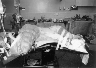
Complications arising from the lateral decubitus position can also be averted with due diligence. We have all placed patients on a bean bag, but the bag must not extend into the axilla of the down arm. A roll (a liter bag of IV solution wrapped in a towel suffices) is placed above the edge of the bean bag just below the axilla. The peroneal nerve in the down leg must be protected with foam and/or gel padding over the fibular head. A pillow is placed between the legs, which are flexed 45 degrees at the hip and knees. The coronal plane of the patient’s thorax must be perpendicular to the floor. Wide tape should be used to secure this position to allow rotation of the bed along its long axis (“airplaning”). Establishing this position assists the surgeon in remaining oriented throughout the procedure, especially during the critical steps of decompressing the spinal cord or placing a vertebral body screw. Some tables are equipped with a compass so that the desired neutral position can be recorded and reset by the anesthesiologist if an “airplane” maneuver is necessary. The perpendicular orientation of the coronal patient plane relative to the floor also allows for more efficient manual reduction of a kyphotic deformity by pressing on the back. The authors routinely “break” or flex the table at the level of the pathology to help open the disc spaces laterally and aid in the insertion of the bone graft (Fig. 146-2). Flexing the table also helps open the space between the 12th rib and the iliac crest. Once the bone graft is in place, the anesthesiologist is asked to return the table to the neutral, unflexed position.


We routinely administer suitable gram-positive antibiotic coverage (e.g., cefazolin 1 g or nafcillin 1 g). In cases of traumatic cord contusion or cord compression caused by tumor, we consider the use of methylprednisolone at least 1 hour before surgery. Using the spinal cord contusion protocol, the patient may receive a bolus of 30 mg/kg over 45 minutes followed by continuous infusion of 5.4 mg/kg per hour for 23 hours.10,11


Paralytic agents are not used after induction to allow for motor response in the event of inadvertent nerve or spinal cord stimulation. The role of somatosensory-evoked potential (SSEP) monitoring is debatable. A decrease in SSEP amplitude of more than 50% and limited or absent intraoperative recovery of amplitude are predictors of a postoperative neurologic deficit.12,13 Despite this reasonable sensitivity and low-false negative rate, SSEP monitoring measures only dorsal column function. False positives are common and often related to anesthetic considerations that can lead to a dangerous desensitization of the surgeon to warnings of intraoperative injury. SSEPs may be useful in deformity cases in which distractive or compressive forces are anticipated and could be reversed.


Motor-evoked potentials may be more accurate than SSEPs in monitoring spinal cord motor function during surgery.14 This technique is extremely sensitive to anesthetics and requires expertise on the part of the anesthesiologist and monitoring team.


Approach and Exposure


The thoracic spine can be approached ventrally by the transmanubrial-transsternal approach, conventional thoracotomy, or thoracoscopic approach. The lumbar spine can be approached by the thoracoabdominal approach, transperitoneal approach, retroperitoneal approach, laparotomy, laparoscopy, balloon-assisted retroperitoneal endoscopy, or low pelvic approaches. These surgical approaches may be performed by the cardiothoracic, general, or vascular surgeon or by the spine surgeon. Detailed preoperative and intraoperative communication about the approach with an approach nonspine surgeon (if used) is important to ensure that not only is the pathologic level exposed but also that the exposure allows the spine surgeon to place instruments perpendicular to the axis of the spine for reconstructive and fixation techniques. Limited exposure may force a screw trajectory in an unsatisfactory cephalad or caudal direction.


Upper Thoracic Spine


Ventral exposure of the rostral levels of the thoracic spine is challenging. The first and second thoracic vertebrae usually can be approached ventrally with a low diagonal or transverse cervical incision. A vertical split of the manubrium often allows exposure down to T3 without sacrificing significant bone. A preoperative sagittal MRI should be obtained and inspected to ensure that the aortic arch does not block ventral access to the T2-3 area. Furthermore, one must be cognizant of the course of the recurrent laryngeal nerve as it emerges dorsal to the brachiocephalic arch to pass between the esophagus and trachea. Although its course is more constant on the left side, low-lying incisions to approach T1 and T2 on the left side put the thoracic duct at risk. This structure is intimately related to the subclavian vein off midline on the left and must be protected. Unrecognized pneumothorax is a complication of this approach because the pleura overlying the medial aspect of the cupola of the lung is adjacent to the spine. Filling the wound with saline and performing a positive pressure inspiration (Valsalva maneuver) at the close of the case is an essential step during closure. An oscillating saw or Gigli saw can be used to remove larger portions of the manubrium, but the retromanubrial space must be palpated to ensure that the brachiocephalic trunk is free. With the patient in the supine position, the upper thoracic spine slopes away from the surgeon, beginning at the T1-2 interspace. It can be difficult to place a ventral plate and screws in this region without a more aggressive removal or splitting of the manubrium or sternum.


Instead of sternotomy, lesions affecting the caudal aspect of T2 to T5 may be approached by a right-sided thoracotomy. The right-sided approach to the upper thoracic spine avoids the aortic arch. One must be cautious, however, to avoid injury to the superior vena cava and supreme intercostal vein. We have found instrumenting the T3-5 area to be easier with a high, right-sided thoracotomy than a midline sternotomy. This experience is particularly true with severe kyphosis (e.g., Scheuermann kyphosis) in this region.


The transaxillary approach is familiar to most vascular surgeons and can be considered for lesions affecting the upper thoracic levels. This approach, however, offers a limited exposure at the base of a cone-shaped cavity and should be reserved for small, more ventrolateral lesions not affecting the entire vertebral body and not requiring (complete) corpectomy or when only open biopsy is necessary. Ventrolateral instrumentation is very difficult because of the limited exposure. The transaxillary approach has associated risks to the lower brachial plexus, long thoracic nerve, and thoracodorsal nerve as well as to vascular structures in the axilla. Splitting of the pectoralis major muscle can also be a source of significant morbidity.


The ventral upper thoracic spine can also be accessed via a third-rib approach in which the patient is positioned in the lateral position with the arm elevated on a rest. The right side is preferable because of the straight course of the brachiocephalic artery. A curved incision is made beginning below the caudal angle of the scapula and ending between the medial scapula and the spinous process of C7. The trapezius and latissimus dorsi muscles are divided medially to minimize denervation, and the scapula is retracted laterally. The dorsal 10 cm of the 2nd, 3rd, and 4th ribs are resected, and the segmental vessels are ligated. The dorsal 3 cm of the 1st rib can also be dissected for additional exposure, but care must be taken not to injure the T1 motor root. The pleura and upper mediastinal structures can then be bluntly dissected for access to the vertebral bodies. Deflation of the lung with a double-lumen endotracheal tube can be very helpful. This approach requires tube thoracostomy placement at the end of the procedure because the parietal pleura is opened and the lung is exposed.


Midthoracic Spine


Lesions involving the midthoracic region are best approached via thoracotomy. Thoracic surgeons are very experienced with this approach. It is recommended that a thoracic surgeon perform the thoracotomy if the spine surgeon is not familiar with this approach. The patient is placed in the lateral decubitus position on a bean bag. The bean bag should not be higher than just below the axilla, and an IV bag or other suitable axillary pad should be placed to protect the brachial plexus and vessels. The area of break in the table should be determined before final positioning so that the desired thoracic level can be placed directly over this area to assist in exposure, opening of the disc spaces, and placement of the graft. Pillows may be placed under the down leg to protect the peroneal nerve and between the legs.


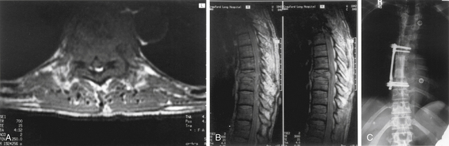
The left side is almost always used for the approach because it is safer and easier to visualize, dissect, and mobilize the aorta and segmental vessels than the vena cava or azygous venous system. It is easier to repair an injured aorta than the vena cava. One should consider obtaining a preoperative axial CT or MRI to assess the location of the aorta. If the aorta is lying very far lateral to the left (Fig. 146-3) or if the pathology is strictly right sided, a right-sided approach can be performed. A standard thoracotomy incision is used beginning approximately two fingerbreadths below the angle of the scapula and coursing ventrally to the midaxillary line. One should select the intercostal space directly over the level of pathology to enter the pleural cavity. We have had satisfactory experience in performing a retropleural thoracotomy. In this procedure, the surgeon separates the endothoracic fascia from the parietal pleura, and the dissection is made down to the rib heads and spine extrapleurally. This is technically more difficult but can obtain a transthoracic approach without the need for a postoperative chest tube. A postoperative radiograph in the recovery room is essential to rule out a significant undetected pneumothorax.


In the lower thoracic spine, this usually corresponds to the rib two numbered levels rostral to the desired vertebral body. For instance, a T8 lesion usually corresponds to the horizontal segment of the 6th rib. Commonly, the rib need not be resected unless it is being harvested for bone graft or if unusually lengthy exposure of the spine is needed. Once the lung is deflated via a double-lumen endotracheal tube and the viscera are packed away with moist towels, the ribs are counted from inside the thoracic cavity. The rib identified as at the same level as the pathology is then exposed in a subpleural fashion down to its insertion on the pedicle. The segmental vessels are identified by blunt or scissor dissection in the midportion of each body. The disc spaces represent the “hills,” and the midvertebral section (where the vessels are located) are the “valleys.” The vessels are ligated with silk suture or metal clips in approximately the midbody. Taking the vessels too close to the aorta risks avulsion during this procedure. Conversely, sacrificing the vessels too close to the neural foramen may interfere with the collateral circulation of the spinal cord. Ligation of the vessels should be performed over the lateral aspect of the vertebral body between the aortic branch point and the neural foramen. Most surgeons and the scoliosis literature agree that up to three adjacent segmental arteries may be taken without neurologic sequelae, but the importance of the artery of Adamkiewicz (T10-L2) is still debated. Some surgeons advocate a preoperative spinal angiogram. Once the vessels are ligated and transected, a subperiosteal dissection of the vertebral body is carried out by using an elevator and unipolar cautery. The anterior longitudinal ligament (ALL) is elevated or incised if ventral release is necessary. If left intact during the exposure, the ALL can serve as a tissue barrier between the aorta and the operative site during the procedure. Some surgeons elevate the ALL sharply or use monopolar cautery from the bone and use that potential space to hold a malleable retractor for added safety.


In anticipation of instrumentation to the levels above and below the pathology, the segmental vessels should be taken here as well. Once this step is complete, a periosteal elevator or monopolar cautery is used to complete a subperiosteal exposure of the diseased level and other levels needed for instrumentation. Thus, the rostral end plate and disc space of the level above and the inferior end plate and disc space of the level below the pathologic levels must be clearly and completely visualized. Ventrally, the exposure is limited by the aorta, but with careful mobilization and retraction (i.e., with large malleable retractors) the cortical bone and disc can be dissected close to (but just short of) the midline. It is critical that a thorough exposure be completed dorsally. The dorsal 2 to 3 cm of each rib (level) involved must be removed with a 1⁄2- or 3⁄4-inch osteotome. Rongeurs or a drill may also be useful. Once the heads of the ribs are disarticulated and removed, the pedicles at each level are exposed and the dorsolateral edge of the vertebral body is confirmed by palpation with a Penfield no. 4. Identification of the pedicle and dorsal vertebral body is essential for recognizing the location of the spinal canal and is necessary for safe and accurate identification of landmarks for placement of instrumentation. Frequently, there is a large mass of soft tissue, including the ligated ends of the segmental vessels, that has been swept into the area of the foramen. One should not attempt to cut away or use the monopolar to cauterize this tissue; the patent segments of the vessels often cause annoying bleeding. Shrinking the tissue near the foramen with the bipolar cautery and then placing a single silk suture through the cauterized mass and sewing it in traction to rib periosteum is often useful. This assists in identifying the spinal canal by moving this tissue in a more dorsal direction. Decompression should not be attempted until the limits of the spinal canal are clearly visualized. Catastrophic neurologic injury may result from initially not identifying accurately the borders of the pedicle, foramen, and dorsal vertebral body (i.e., the spinal canal).

Stay updated, free articles. Join our Telegram channel

Aug 31, 2016 | Posted by in NEUROLOGY | Comments Off on Ventral and Lateral Thoracic and Lumbar Fixation Techniques

Full access? Get Clinical Tree

Get Clinical Tree app for offline access