Ventricular system
The central nervous system (CNS) contains an interconnecting series of chambers and channels that are derived from the lumen of the embryonic neural tube. In the spinal cord, this is represented by the vestigial and insignificant central canal. Within the brain, however, the enormous growth and distortion of the original tube-like structure is paralleled by the development of an elaborate series of chambers, the cerebral ventricles (Figs 6.1, 6.2, see also Fig. 1.22).


Topographical anatomy of the ventricular system
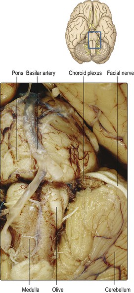
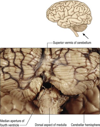
Passing rostrally from the spinal cord to the brainstem, the central canal of the spinal cord moves progressively more dorsal until, in the rostral (open) medulla, it opens out into a wide and shallow depression, the fourth ventricle (Fig. 6.3), which lies on the dorsal surface of the brainstem beneath the cerebellum. The fourth ventricle is rhomboid or diamond-shaped. On each side, a lateral recess extends towards the lateral margin of the brainstem and is in continuity, through a small lateral aperture (the foramen of Luschka), with the subarachnoid space of the cerebellopontine angle (Fig. 6.4). For the most part, the roof of the fourth ventricle is formed by the cerebellum. However, the roof of the caudal part consists of pia and ependyma, a central defect in which constitutes the median aperture of the fourth ventricle, or the foramen of Magendie. The latter provides communication between the lumen of the ventricle and the cisterna magna of the subarachnoid space (Fig. 6.5). The rostral part of the roof of the fourth ventricle is partly formed by the superior cerebellar peduncles on either side, the space between them being bridged by the thin superior medullary velum.

The fourth ventricle extends rostrally as far as the pontomesencephalic junction, where it becomes continuous with the cerebral aqueduct. The cerebral aqueduct is a narrow channel which passes throughout the length of the midbrain, beneath the inferior and superior colliculi. The simple, tubular configuration of the cerebral aqueduct, in comparison to the rest of the cerebral ventricular system, reflects the relatively undifferentiated nature of the midbrain in comparison to the forebrain. At the rostral margin of the midbrain, the cerebral aqueduct opens into the third ventricle. This is a midline, narrow, slit-like cavity, the lateral walls of which are formed by the thalamus and hypothalamus on either side (Fig. 6.2 and see Fig. 12.2). The roof of the third ventricle is formed by pia-ependyma, which spans between two nerve fibre bundles, the striae medullaris thalami, which are situated along the dorsomedial border of the thalamus. In the rostral part of the third ventricle lies an aperture, the interventricular foramen, or foramen of Monro, which is located between the column of the fornix and the anterior pole of the thalamus.
The interventricular foramen provides communication, on either side, with the extensive lateral ventricle located within the cerebral hemisphere (Fig. 6.6 and see Fig. 16.15). The lateral ventricle is approximately C-shaped. It consists of an anterior (frontal) horn, a body, posterior (occipital) horn and an inferior (temporal) horn. The anterior horn of the lateral ventricle is that part lying anterior to the interventricular foramen. Its lateral wall is formed by the head of the caudate nucleus and its roof is the corpus callosum (see Figs 13.3–13.7). The medial wall is formed by the septum pellucidum. This thin sheet spans between the corpus callosum and fornix in the midline and separates the anterior horns of the two lateral ventricles. The body of the lateral ventricle extends behind the interventricular foramen, having as its floor the thalamus and tail of the caudate nucleus. More posteriorly, the small posterior horn extends towards the occipital pole but the principal course of the ventricle sweeps downwards and forwards to form the extensive inferior horn, which lies in the temporal lobe. In the floor of the inferior horn lies the hippocampus, while in its roof runs the much attenuated tail of the caudate nucleus (see Figs 13.9–13.12 and 16.15).
Related posts:

Stay updated, free articles. Join our Telegram channel

Full access? Get Clinical Tree


