♦ Preoperative
Operative Planning
- Magnetic resonance imaging and/or computed tomography (CT) scan to review ventricular size and associated intracranial anatomy
- Stereotactic guidance for intraventricular shunt placement may be helpful in small ventricles
- Site of burr hole and subsequent ventricular catheter placement is usually determined by ventricular anatomy, with preference toward side of larger ventricle or side opposite dominant hemisphere
- Frontal or parietooccipital placement is determined by ventricular anatomy and surgeon preference
- Site of distal peritoneal catheter is usually same side as the ventricular catheter unless previous surgeries or other contraindications favor opposite side
- Basic shunt tray
- Intraventricular catheter; antibiotic-impregnated catheters have been shown to decrease incidence of shunt infections
- Shunt valve; choice and setting is usually case specific and surgeon dependent
- Tunneler
- Pleural catheter
♦ Intraoperative
- Antibiotics are given prior to incision.
Positioning
- Frontal intraventricular catheter placement
- Patient is supine with the head in the neutral position on a doughnut pillow or, when neuronavigation is used, in three-pin fixation
- Head of bed elevated 30 degrees to prevent air entry and excessive CSF loss
- Patient is supine with the head in the neutral position on a doughnut pillow or, when neuronavigation is used, in three-pin fixation
- Parietal intraventricular catheter placement
- Patient is supine with head on doughnut pillow, horseshoe, or in three-pin fixation when neuronavigation is used
- Head turned 90 degrees to opposite side, with burr hole site up
- Shuolder roll placed under burr hole side to slightly extend neck and allow for smoother passage of the tunneler
- Eyes taped shut
- Proper padding of all pressure points
- Patient is supine with head on doughnut pillow, horseshoe, or in three-pin fixation when neuronavigation is used
Planning of Sterile Scrub and Preparation
- Entire operative field shuold be prepped and exposed, including area over neck and chest where the tunneler will be passed.
- Shave is surgeon dependent; minimal shave includes area around incision. Shunt tract path is often also shaved.
- Betadine scrub and paint
- 3M drapes and/or sterile towels placed along the exposed head, neck, thorax, and abdomen, and Ioban placed over draped area
Incision
- Cranial incision
- For frontal burr holes, curvilinear or linear incision shuold be made over Kocher’s point.
- For parietooccipital burr hole, curvilinear or linear incision is made approximately three fingerbreadths behind the ear and three fingerbreadths above the ear in adults, or along the flat portion of the parietal boss in children.
- For frontal burr holes, curvilinear or linear incision shuold be made over Kocher’s point.
- Thoracic incision
- Linear incision along the anterior axillary line, within one of the intercostal spaces between the 4th to 6th ribs, is most convenient and preferred (Fig. 81.1A).
< div class='tao-gold-member'>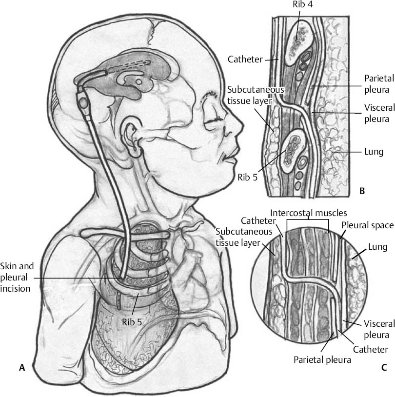 Only gold members can continue reading. Log In or Register to continue
- Linear incision along the anterior axillary line, within one of the intercostal spaces between the 4th to 6th ribs, is most convenient and preferred (Fig. 81.1A).


