♦ Preoperative
Operative Planning
- Magnetic resonance imaging and computed tomography (CT) scan to review ventricular size and associated intracranial anatomy
- CT-guided stereotactic catheter placement may be indicated for intraventricular shunt placement in small ventricles.
- Site of burr hole and subsequent ventricular catheter placement is usually determined by ventricular anatomy, with preference toward side of larger ventricle or site opposite dominant hemisphere.
- Frontal or parietooccipital placement is determined by ventricular anatomy and surgeon preference.
- Site of distal peritoneal catheter is usually same side as the ventricular catheter unless previous surgeries or other contraindications favor opposite side.
Special Equipment
- Basic shunt tray
- Intraventricular catheter; antibiotic impregnated catheters have been shown to decrease incidence of shunt infections
- Shunt valve; choice and setting is usually case specific and surgeon dependent
- Tunneler
- Peritoneal catheter
Intraoperative
- Antibiotics are given within 30 minutes prior to incision
Positioning
- Frontal intraventricular catheter placement
- Patient is supine with the head in the neutral position on a doughnut pillow or, when neuronavigation is used, in three-pin fixation
- Head of bed elevated 30 degrees to prevent air entry and excessive CSF loss
- Patient is supine with the head in the neutral position on a doughnut pillow or, when neuronavigation is used, in three-pin fixation
- Parietal intraventricular catheter placement
- Patient is supine with head on doughnut pillow, horseshoe, or in three-pin fixation when neuronavigation is used
- Head turned 90 degrees to opposite side, with burr hole site up
- Shoulder roll placed under burr hole side to slightly extend neck and allow for smoother passage of the tunneler
- Eyes taped shut
- Proper padding of all pressure points
- Patient is supine with head on doughnut pillow, horseshoe, or in three-pin fixation when neuronavigation is used
Sterile Scrub and Prep
- Entire operative field should be prepped and exposed, including area over neck and chest where the tunneler will be passed.
- Shave is surgeon dependent; minimal shave includes area around incision. Shunt tract path is often also shaved.
- Betadine scrub and paint
- 3M drapes and/or sterile towels placed along the exposed head, neck, thorax and abdomen, and Ioban (3M) placed over draped area
Planning of Incision
- For frontal burr holes, curvilinear or linear incision should be made over Kocher’s point
- For parietooccipital burr hole, curvilinear or linear incision is made approximately three fingerbreadths behind the ear and three fingerbreadths above the ear in adults, or along the flat portion of the parietal boss in children (Fig. 78.1)
- Abdominal incision: horizontal linear incision usually in lateral quadrant, approximately two fingerbreadths below rib cage
- Bipolar cautery for hemostasis
- Small, self retaining retractor
Burr Hole
- Retractor pulled caudally or laterally to expose burr hole site and ensure that no hardware lies under suture line
- Burr hole is made with perforator, hand twist drill, or knife and Kerrison punch in children, or with air-powered perforator in adults
< div class='tao-gold-member'>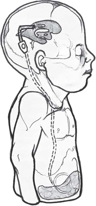 Only gold members can continue reading. Log In or Register to continue
Related posts:

Stay updated, free articles. Join our Telegram channel


Full access? Get Clinical Tree


