♦ Preoperative
Operative Planning
- Computed tomography (CT) scan or magnetic resonance imaging to review ventricular size and associated intracranial anatomy
- Prior head CT when patient’s shunt was known to be working is useful in helping to determine shunt failure via comparison of ventricular size
- Neuronavigation may be indicated for intraventricular shunt placement in small ventricles
- Shunt series to evaluate for any kinks or disconnection in the shunt system
- Clinical assessment with palpation of shunt bulb and shunt tapping when indicated to assist in determining site of shunt failure
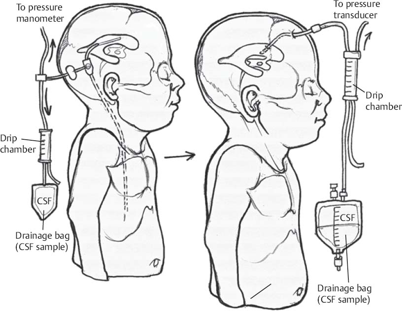
Special Equipment
- Basic shunt tray
- Intraventricular catheter; antibiotic-impregnated catheters have been shown to decrease incidence of shunt infections
- Shunt valve; choice and setting is usually case specific and surgeon dependent
- Tunneler
- Distal catheter
- A 10-mL saline filled syringe attached to manometer with stopcock and a blunt tip needle
- If problem is related to the peritoneal cavity (i.e., fluid loculation secondary to adhesions), consider general surgery consult for intraoperative assistance at distal catheter fragment removal and replacement.
Only gold members can continue reading. Log In or Register to continue


