Loss of Vision
Visual loss may be due to a problem within the eye, the optic nerve or the posterior visual pathways.
Local Eye Disease
- Disturbance of the eye and vision is common and it is important to consider local changes within the eye.
- Consider corneal damage, cataract, refractive errors, glaucoma and retinal disturbance.
- Correct a refractive error producing myopia with a pinhole early in the course of considering the cause of visual loss. Having excluded the causes of visual disturbance from the surface of the eye back to the retina, consider neurological dysfunction of the optic nerve or posterior visual pathways.
Optic Nerve Disturbance
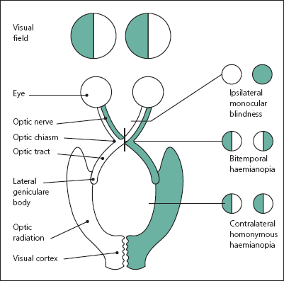
- Optic nerve lesions cause a disturbance of central vision (scotoma) with a reduction in visual acuity (Figure 7.1).
- Visual loss due to anterior lesions of the optic nerve reduces visual acuity leading to reduced perception of distant objects and potential difficulty with reading.
- Determine the loss of acuity: test in either eye separately for central vision using, for example, a Snellen chart.
- Understanding the temporal onset of central visual loss is often the most useful determinant in recognising the aetiology of anterior optic nerve disturbance.
- Determining whether central visual loss is episodic (and if so, of what duration) or persistent is further discriminating.
Optic Nerve Disturbance Causing Sudden Onset of Transient Visual Loss
- Sudden onset of unilateral visual loss may be vascular in origin due to disturbance in the ophthalmic artery (amaurosis fugax). Often a falling or rising curtain affecting either the upper or lower half of the vision from one eye due to occlusion of either the superior or the inferior ophthalmic artery is described. Hence, so-called altitudinal defects often reflect a vascular aetiology.
- Amaurosis fugax is a transient ischaemic episode of the ophthalmic artery and typically occurs in older patients with vascular risk factors.
- Sudden temporary central visual loss also occurs with so-called obscurations of vision secondary to raised intracranial pressure.
- Such visual obscurations are typically very brief, lasting seconds to minutes only. Visual obscurations are normally seen where background intracranial pressure is high, for whatever reason, and then with a provoking manoeuvre (such as coughing or straining) which raises intracranial pressure further causing brief visual loss. With visual obscurations, look for swollen discs.
- Transient episodic visual loss can occur with previous optic neuritis (demyelination/multiple sclerosis) when the patient’s body temperature rises, such as with exercise, the so-called Uhthoff’s phenomena.
Optic Nerve Disturbance Causing Persistent Visual Loss
- Persistent visual loss may be acute, subacute or chronic in its progression.
- Acute (occurring over minutes) persistent visual loss is often due to a vascular aetiology affecting the ophthalmic arteries or veins.
- Subacute relapsing remitting central visual loss is commonly due to inflammation/demyelination within the optic nerves, such as is seen in multiple sclerosis.
- Subacute and chronic progressive central visual loss may be due to a compressive structural cause and requires urgent MRI imaging. Lesions compressing the optic nerve are frequently benign but prompt intervention remains important to preserve vision.
- If a structural cause has been ruled out, toxic (tobacco/alcohol), nutritional (B1/B12), drug-induced and hereditary optic neuropathies (Leber’s optic neuropathy) should be considered.
Only gold members can continue reading. Log In or Register to continue


