Visual Field Examination
PURPOSE
The purpose of the visual field examination is to assess the function of the visual pathway that begins in the eyes and ends in the occipital cortex, because lesions located along different regions of this pathway produce characteristic visual field abnormalities.
WHEN TO EXAMINE THE VISUAL FIELDS
Confrontational visual field testing is a quick and easy way of discovering significant visual field loss, and it should be performed on all patients as part of a standard neurologic examination.
NEUROANATOMY OF THE VISUAL FIELDS
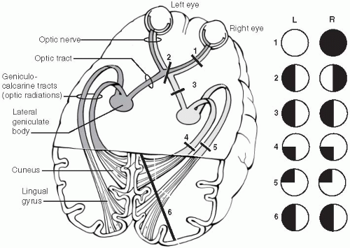
The visual pathway is pictured in Figure 13-1. The nasal (medial) part of each retina sees the temporal visual world, and the temporal (lateral) part of each retina sees the nasal visual world. Visual information from each retina travels through the optic nerves into the optic chiasm. At the optic chiasm, the visual information from the nasal part of each retina crosses to the other side and continues as the optic tract, whereas the visual information from the lateral part of each retina remains uncrossed, also continuing as the optic tract. Each optic tract synapses in the lateral geniculate nucleus. From the lateral geniculate nuclei, the visual information continues onward toward the occipital cortex as the optic radiations. Visual information from the lower retina (which sees the upper fields) travels through the optic radiations that are located in the temporal lobes, reaching the lower occipital cortex. Visual information from the upper retina (which sees the lower fields) travels through the optic radiations that are located in the parietal lobes, reaching the upper part of the occipital cortex.
EQUIPMENT NEEDED FOR THE VISUAL FIELD EXAMINATION
None.
HOW TO EXAMINE THE VISUAL FIELDS
Stand a few feet in front of the patient, with your head at approximately the same level as the patient, looking directly at the patient’s eyes.
Instruct the patient to look at your nose throughout the examination, and have the patient cover one eye with his or her hand. Ask the patient to count the total number of fingers you’ll be holding up.
Check the visual fields by holding up one, two, or five fingers in the vertical plane that is just between you and the patient, checking each of the four quadrants. Test at least four separate areas: the left and right upper visual fields and the left and right lower visual fields. You do not need to hold the hands far into the periphery, only approximately 1 ft away from the midline. In most cases, you can quickly check both upper fields at the same time (for example, by holding up one finger with your left hand and
two fingers with your right hand, asking the patient to tell you the total number of fingers you’re holding up), and then examine both lower fields at the same time. If this is confusing to the patient, check each quadrant separately.

Figure 13-1 Illustration of the visual pathway from the eyes to the occipital cortex. The characteristic visual field deficits that would occur due to each of the numbered lesions at various regions of the visual pathway are shown. See text for further details. (Redrawn from Gilman S, Newman SW. Vision. In: Manter and Gatz’s essentials of clinical neuroanatomy and neurophysiology, 8th ed. Philadelphia: FA Davis Co, 1992:196.)
Stay updated, free articles. Join our Telegram channel

Full access? Get Clinical Tree

 Get Clinical Tree app for offline access


