Visual system
Vision is the most highly developed and versatile of all the sensory modalities and, arguably, the one on which humans are most dependent. The optic nerve and retina develop from the prosencephalic primary brain vesicle and, therefore, are regarded as an outgrowth of the brain itself. Vision commences with the formation of an image of the external world on the photoreceptive retina. The retina encodes visual information in the discharge of neurones that project to the brain through the optic nerve. Fibres of the optic nerve undergo hemidecussation in the optic chiasm and project to the lateral geniculate nucleus of the thalamus. Thalamocortical neurones in turn project to the primary visual cortex in the occipital lobe of the cerebral hemisphere, where visual perception occurs.
The eye
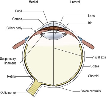
In the eye, the eyeball, or globe, is approximately spherical in shape (Fig. 15.1). Near its posterior pole emerges the optic nerve. The eyeball may be considered to consist of three concentric layers of tissue, the outermost of which is tough, fibrous and protective. Over most of the globe it forms an opaque white coat, the sclera, to which are attached the extraocular muscles that move the eyeball. Over the anterior pole of the globe it forms the transparent cornea, through which light enters the eye.
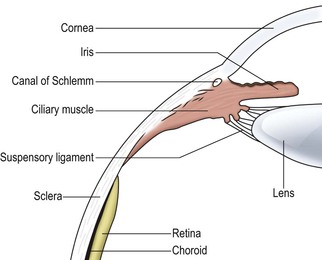
Near to the anterior margin of the sclera, two rings of smooth muscle extend into the lumen of the eyeball (Fig. 15.2). The most anterior of these is the iris, which has a central aperture, the pupil, through which light is admitted to the posterior part of the eye. Some of the muscle fibres of the iris are arranged in a circular fashion, while others are oriented radially. They are under the control of the autonomic nervous system. Circular fibres are innervated by postganglionic parasympathetic neurones, which act to constrict the pupil and reduce the amount of light falling upon the retina (see p. 105). Radial fibres are innervated by postganglionic sympathetic neurones which dilate the pupil.
Behind the iris lies the ciliary body containing ciliary muscle, which receives innervation from the parasympathetic nervous system. The central aperture within the annulus of the ciliary body is occupied by the transparent, biconvex lens, which focuses light upon the retina. The lens is held in place by a suspensory ligament that is attached to the peripheral margin of the lens and to the ciliary body. Contraction of the ciliary muscle alters the shape and, therefore, the focusing power (focal length) of the lens, a process known as accommodation. The lens and suspensory ligament divide the lumen of the eyeball into an anterior and a posterior part. The anterior part, in front of the lens, contains a thin, watery fluid, aqueous humour, which is continuously secreted from the ciliary body. It is also reabsorbed into the ciliary body where it is drained by a small duct, the canal of Schlemm, through which it is returned to the venous system. The posterior part of the globe contains a gelatinous material known as vitreous humour. Behind the ciliary body, the inner surface of the sclera is lined by the choroid, the cells of which contain dark pigment that absorbs light and thus reduces reflection within the eye. Lining the inner surface of the choroid is the photoreceptive retina.
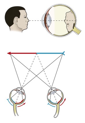
Light passes from objects in the field of vision (visual field), through the narrow aperture of the pupil to subtend an image upon the retina. An object in the visual field, upon which attention is focused, subtends an image that is centred near the posterior pole of the eye along the line of the visual axis (Fig. 15.1). At this point, which is known as the fovea centralis, and the surrounding 1 cm which is known as the macula lutea, the retina is specially modified for maximal visual acuity (resolving power). The basic optical properties of the eye, which may be likened to those of a pinhole camera, dictate that the image so formed is inverted in both lateral and vertical dimensions (Fig. 15.3). Furthermore, objects that lie in the left half of the visual field form an image upon the nasal (right) half of the left retina and the temporal (right) half of the right retina, and vice versa. Medial to the macula is a region where retinal axons accumulate to leave the eye in the optic nerve. This is known as the optic disc. Photoreceptors are absent from this region, which is also, therefore, referred to as the blind spot.

< div class='tao-gold-member'>
Stay updated, free articles. Join our Telegram channel

Full access? Get Clinical Tree


