Fig. 1
Comparison of brain volume determination between those directly measured versus reconstitution after the brain edema procedure; SEM standard error of the mean; n = 10/group

Fig. 2
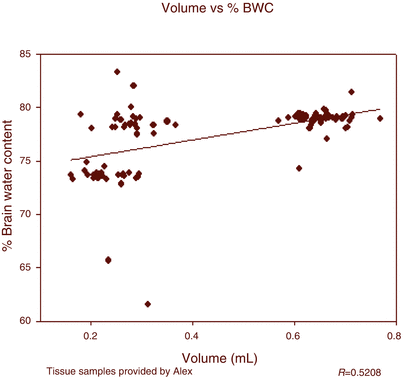
Demonstration of correlation between percentage brain water content and volume. Note fold changes in volume for each percentage edema. Left cluster infratentorial, right cluster supratentorial (i.e., cerebral hemispheres)

Fig. 3
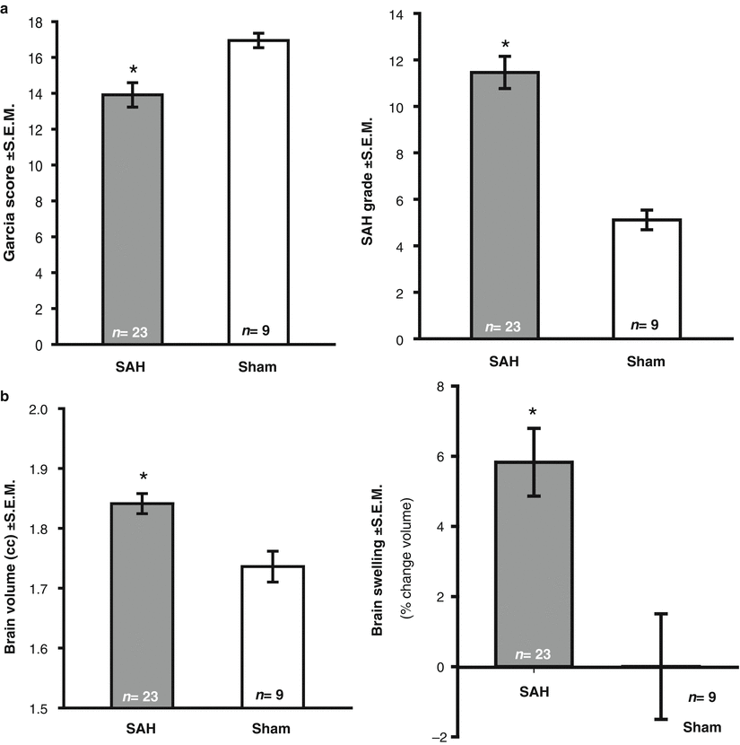
(a) Neurological deficit (Garcia score; left panel), SAH grade (right panel) measured following SAH; (b) calculated brain volume (left panel), and mathematical conversion into percentage swelling (right panel) measured following SAH; (asterisk) <0.05 versus sham; SEM standard error of mean; n = 9–23/group
Conclusion
Translational stroke studies, including animal modeling, are greatly needed to safely integrate basic preclinical scientific principles ahead of clinical application [32–36]. Thus, this modification of the “wet-dry” brain edema method permits brain volume determination using valuable post hoc dried brain tissue. Perhaps the greatest strength of this approach is the acquisition of additional valuable data without greatly added expense to neuroscience laboratories. Nonetheless, the application of these volumetric measurements needs validation in other studies, across models and species. In the meantime, this breakthrough technique remains a hammer without a nail but we hope not for much longer.
Acknowledgment
This study was partially supported by the National Institutes of Health grant RO1 NS078755 (Dr Zhang).
Disclosures
None
References
1.
Keep RF, Hua Y, Xi G (2012) Brain water content. A misunderstood measurement? Transl Stroke Res 3:263–265PubMedCentralCrossRefPubMed
2.
Hasegawa Y, Nakagawa T, Uekawa K, Ma M, Lin B, Kusaka H, Katayama T, Sueta D, Toyama K, Koibuchi N, Kim-Mitsuyama S (2014) Therapy with the combination of amlodipine and irbesartan has persistent preventative effects on stroke onset associated with BDNF preservation on cerebral vessels in hypertensive rats. Transl Stroke Res. doi:10.1007/s12975-014-0383-5 PubMed
3.
Schlunk F, Schulz E, Lauer A, Yigitkanli K, Pfeilschifter W, Steinmetz H, Lo EH, Foerch C (2014) Warfarin pretreatment reduces cell death and MMP-9 activity in experimental intracerebral hemorrhage. Transl Stroke Res. doi:10.1007/s12975-014-0377-3 PubMed
4.
Chen Q, Zhang J, Guo J, Tang J, Tao Y, Li L, Feng H, Chen Z (2014) Chronic hydrocephalus and perihematomal tissue injury developed in a rat model of intracerebral hemorrhage with ventricular extension. Transl Stroke Res. doi:10.1007/s12975-014-0367-5
5.
6.
Li Q, Khatibi N, Zhang JH (2014) Vascular neural network: the importance of vein drainage in stroke. Transl Stroke Res 5:163–166PubMedCentralCrossRefPubMed
7.
8.
Hoda MN, Bhatia K, Hafez SS, Johnson MH, Siddiqui S, Ergul A, Zaidi SK, Fagan SC, Hess DC (2014) Remote ischemic perconditioning is effective after embolic stroke in ovariectomized female mice. Transl Stroke Res 5:484–490PubMedCentralCrossRefPubMed
9.
Khanna A, Kahle KT, Walcott BP, Gerzanich V, Simard JM (2014) Disruption of ion homeostasis in the neurogliovascular unit underlies the pathogenesis of ischemic cerebral edema. Transl Stroke Res 5:3–16PubMedCentralCrossRefPubMed
Stay updated, free articles. Join our Telegram channel

Full access? Get Clinical Tree


