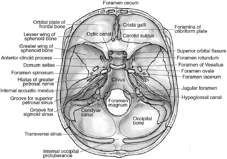
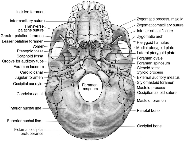
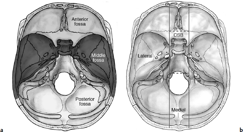
2 Anatomy of the Skull Base and Related Structures: Elements of Surgical Anatomy The skull has two components: Neurocranium (“braincase”): houses the brain. Splanchnocranium (or viscerocranium): formed by the bones of the face. The neurocranium has two components: Calvaria (“skull cap”) Skull base (basicranium): the floor of the cranial cavity, on which the brain rests. The skull base has two surfaces: Endocranial (inner): the floor of the cranial cavity, on which the brain rests (Fig. 2.1). Exocranial (external) surface (Fig. 2.2). • The bones, which form the skull base, are the frontal, sphenoid, ethmoid, temporal, and occipital bones (the anterior part of the exocranial surface is also formed by the zygomatic, maxillary, and palatine bones). • The inner surface of the skull base can be divided into three transverse parts (anterior, middle, and posterior fossae) (Fig. 2.3a) and in three sagittal parts (central and lateral parts) (Fig. 2.3b). • The median part of the anterior skull base covers the upper nasal cavity and the sphenoid sinus; the middle part contains the cavernous sinuses laterally, which house the carotid arteries (parasellar compartments); the posterior part includes the clivus, which reaches the anterior margin of the great occipital foramen. Fig. 2.1 Endocranial surface of the skull base. Fig. 2.2 Exocranial surface of the skull base. Fig. 2.3a,b (a) Skull base: anterior, middle, and posterior cranial fossae. (b) Lateral and median regions. CSB, central skull base (see also Fig. 5.7 on page 114). • Anterior skull base: formed by one ethmoidal, two frontal, and one sphenoid bones. The anterior skull base (ASB) is delimited anteriorly by the frontal bone and the posterior wall of the frontal sinus, and posteriorly by the lesser wings of the sphenoid bone. The lateral parts of the anterior skull base form the roof of the orbits, on which the frontal lobes of the brain lie. The median (central) part is formed by the crista galli, the cribriform plate of the ethmoid bone, and the planum of the sphenoid bone. • Middle skull base: formed by one sphenoid and two temporal bones. The middle skull base (MSB) is delimited anteriorly by the lesser wings of the sphenoid bones, and posteriorly by the surface of the petrous part of the temporal bone. The temporal lobe lies in the middle cranial fossa. The central part of the middle cranial fossa is defined as the sella turcica (part of the sphenoid bone). The sellar compartment contains the hypophysis. It is separated from the suprasellar compartment (brain) by a meningeal sheet, the diaphragma sellae, which has an opening at the center called the ostium of the diaphragma sellae. The pituitary stalk crosses through this opening. The lateral parts of the sellar compartments are defined as “parasellar compartments.” This is the location of the cavernous sinus, which is crossed by the cranial nerve (CN) VI and the internal carotid artery (ICA). The lateral wall of the cavernous sinus is also lined by CN III, IV, V1, and V2. For each skull base foramen, it is very important to remember its neurovascular relationships. Tables 2.1 and 2.2 summarize the foramina of the endocranial surface and the exocranial surface of the skull base with its contents, respectively. Table 2.3 summarizes the foramina and other structures visible on the splanchnocranium (Fig. 2.4). • Posterior skull base: formed by the occipital and temporal bones. This fossa is delimited anteriorly by the posterior walls of the petrous bone and posteriorly by the grooves for the transverse sinuses. It contains the foramen magnum, in which the medulla oblongata continues downward into the spinal cord. • The pituitary fossa is in the central part of the sphenoid bone, at the center of the skull. The sellar region is delimitated anteriorly by the jugum sphenoidale, which is the most posterior border of the planum sphenoidale, anterolaterally by the extension of the lesser sphenoid wings (anterior clinoid processes, ACPs), laterally by the greater sphenoid wings, posterolaterally by the posterior clinoid processes (PCPs) and petrous apex, and posteriorly by the clivus. • The ACP forms the anterolateral bony protuberance; the optic canal runs medial to it. The optic strut is the medial attachment of the ACP, forming part of the optic canal and separating the carotid sulcus from the optic canal. • The portion of the ICA passing between the optic strut and the superior surface of the ACP is the clinoidal segment of the ICA.1 • Chiasmatic groove (sulcus): a depression between the tuberculum sellae, the planum sphenoidale, and the optic foramina. • Anterolaterally to the sella and ACP lie the superior orbital fissures on each side. The cavernous sinus extends from the petrous apex to the superior orbital fissure (SOF).2 • The normal average size of the pituitary fossa is 13 × 17 × 15 mm, with an average volume of 1,100 mm3. Surgical Anatomy Pearl An anatomic variant is the presence of the middle clinoid process, which can bridge the ACP. In such a situation, the carotid artery would run through a caroticoclinoid foramen. Table 2.1 Foramina of the Skull Base: Endocranial Surface and Its Contents
 Skull Definitions
 2.1 Skull Base Anatomy—Anterior and Middle Skull Base
Osteology of the Cranial Fossae
Sellar and Parasellar Compartments
Foramen cecum | Emissary vein from the superior sagittal sinus to frontal sinus and nose; eventually anterior falcine artery |
Foramina of the cribriform plate | Olfactory nerve bundles from the nasal mucosa to the olfactory bulb |
Anterior, middle (variable), and posterior ethmoidal foramina | Anterior, middle (whenever present the middle foramina) and posterior ethmoid arteries and veins |
Optic canal | CN II and ophthalmic artery |
Superior orbital fissure | CNs III, IV, VI, V1, superior and inferior ophthalmic veins, orbital branches of the MMA, sympathetic fibers (that enter mainly with nasociliary nerve), parasymphathetic fibers (entering with CN III) |
Inferior orbital fissure | Venous channels connecting orbital venous system with pterygoid plexus, small nameless arterial branches, branches from pterygo palatine ganglion and zygomatic nerve from infraorbital nerve |
Foramen rotundum | CN V2, accompanied by some emissary veins and an arterious branch from the internal maxillary artery |
Foramen ovale | CN V3, lesser superficial petrosal nerve, accessory meningeal artery branch from the maxillary artery |
Foramen spinosum | Middle meningeal artery and vein, meningeal recurrent branch of V3 |
Foramen lacerum | Meningeal branches from the ascending pharyngeal artery, as well as the nerve of pterygoid canal, cartilage |
Vidian canal | Vidian nerve (formed by the GSPN and the deep petrosal nerve), veins, and two arteries |
Hiatus for greater petrosal foramen | Greater superior petrosal nerve |
Hiatus for lesser petrosal nerve | Lesser superior petrosal nerve |
Internal acoustic canal | CNs VII–VIII, labyrinthine artery |
Jugular foramen | CNs IX, X, XI, convergence of inferior petrosal sinus and sigmoid sinus into the internal jugular vein, Jacobson’s nerve (tympanic branch of CN IX), Arnold’s nerve (auricular branch of CN X), posterior meningeal artery (from the VA) |
Hypoglossal canal | CN XII with meningeal artery |
Foramen magnum | Medulla oblongata, vertebral arteries, spinal roots of CN XI, anterior spinal artery, posterior spinal arteries, and posterior meningeal arteries |
Abbreviations: CN, cranial nerve; GSPN, greater superficial petrosal nerve; MMA, middle meningeal artery; VA, vertebral artery.
Table 2.2 Foramina of the Exocranial Surface of the Skull Base
Carotid canal | ICA surrounded by the sympathetic plexus |
Greater palatine foramen | Greater palatine nerve (from V2) and vessels |
Lesser palatine foramen | Lesser palatine nerve (from V2) and vessels |
Sphenopalatine foramen | Nasopalatine nerve (from V2), nasal nerve (from V2), sphenopalatine artery |
Stylomastoid foramen | CN VII |
Foramen magnum | See Table 2.1 |
Petrotympanic fissure | Anterior tympanic branch of the internal maxillary artery |
Abbreviation: ICA, internal carotid artery.
Table 2.3 Foramina of the Splanchnocranium
Supraorbital foramen/incisura | Supraorbital nerve (from V1), supraorbital vein and artery |
Infraorbital canal and foramen | Infraorbital nerve (from V2), infraorbital vein and artery |
Zygomaticotemporal foramen | Zygomaticotemporal nerve and vessels |
Zygomaticofacial foramen | Zygomaticofacial nerve and vessels |
Incisive foramen | Nasopalatine nerve (from V2) and vessels |
Mandibular foramen | Inferior alveolar nerve (from V3) |
Mental foramen | Mental nerve (from V3) |
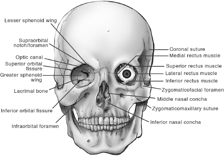
Fig. 2.4 Anatomic structures of the splanchnocranium and orbits.
The parasellar region encompasses the anatomic compartments around the sella:
Parasellar compartments: cavernous sinuses
Suprasellar compartment: region above the diaphragma sella, with the suprasellar cistern and its contents
Retrosellar compartment: clivus
• The pituitary fossa contains the pituitary gland. The pituitary gland is formed by an anterior part (adenohypophysis), a more orange-colored posterior lobe (neurohypophysis), and an often cystic intermediate lobe.
 The anterior lobe forms the pars tuberalis at the lower part of the pituitary stalk.
 The inferior surface of the gland conforms to the shape of the sellar floor.
 The superior part can be more flat or concave around the stalk. The stalk crosses the diaphragma into the ostium of the diaphragma sellae.
Cavernous Sinuses
The cavernous sinuses are lateral to the sphenoid sinus, sella turcica, and pituitary gland (Fig. 2.5). Each cavernous sinus sits in the central aspect of the middle fossa and is lateral to the sella.
• Cavernous sinus (CS) is the historical definition of the lateral sellar compartment; it is part of the extradural neural axis compartment (EDNAC), which extends from the coccyx to the orbit.2 The name incorrectly suggests that the CS is a “sinus,” but it is not a sinus like the superior sagittal sinus; rather, it is a multiloculated network of lacunae on the lateral compartment of the sellar region.
• The CS is one component of the basal venous system, which consists of a valveless network of interconnected venous channels. Should an obstruction occur in one region, venous flow can decant into a collateral system and so allow venous drainage. These veins include anterior and posterior intercavernous sinuses, the petroclival plexus, the superior and inferior ophthalmic veins, the sphenoparietal sinus, the superior and inferior petrosal sinuses, a variety of emissary veins that can enlarge in the event of occlusion, and some variable veins from the brain (e.g., vena Vasaliana).
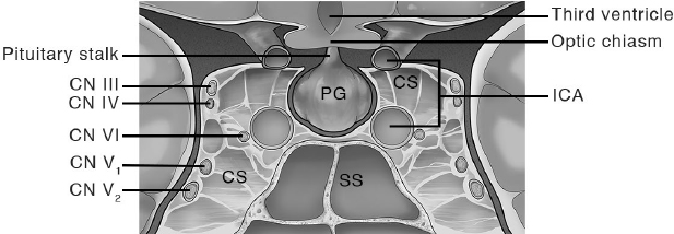
Fig. 2.5 Cavernous sinus (CS), coronal section. ICA, internal carotid artery; SS, sphenoidal sinus.
Anatomic Boundaries: Dural Layers of the Cavernous Sinus
• Lateral wall: A double layer of dural mater, the outer layer is continuous with the internal layer of the dura of the middle cranial fossa and tentorium, whereas the inner layer of the lateral wall is formed by the epineurium of CNs III, IV, and V. The outer layer of the middle fossa dura separates at the lateral margin of the cavernous sinus to continue as the periosteum and to form the floor and medial wall of the cavernous sinus. There is a cleavage plane between the medial and lateral layers, which may be dissected without entering the venous compartment of the CS3 (see Chapter 21).
• Medial wall: layer of dura/periosteum.
• Roof: The third or distal ring of dura around the ICA forms the carotico-oculomotor membrane.4,5 The clinoid and carotid triangles are part of the roof as well. The roof of the CS has the shape of a trapezium, extending from the diaphragma sellae medially, to the anterior petroclinoid ligament laterally, from the base of the anterior clinoid process anteriorly, and to the posterior petroclinoid ligament posteriorly.3 It provides a base for the cistern of the CN III.
• Posterior wall: dura of the clivus.
• Contents of the double layer of the lateral wall: CNs III and IV and the branches of the trigeminal nerve run within the double layer.
• Contents of the cavernous sinus: venous lacunae, CN VI, fat, cavernous ICA surrounded by sympathetic plexus, with its branches:
 Meningohypophyseal trunk: Branches of this trunk are variable, but in pure form consist of the tentorial artery (also known as the artery of Bernasconi-Cassinari), dorsal meningeal artery (also known as the clival artery), and the inferior hypophyseal artery.
 Inferolateral trunk: branches supplying the intracavernous nerves.
 Capsular artery: McConnel’s artery, present in 30% of cases.6,7
 Ophthalmic artery: generally extracavernous, but in 3 to 8% it originates from the infraclinoidal segment of the ICA.8
Cranial nerve VI runs from the posterior part of the CS, passing through the Dorello canal, medially to the Meckel’s cave and medial to the trigeminal root,9 to the SOF, medial and superior to V1.
Triangles of the Skull Base
Specific anatomic landmarks create triangular-shaped corridors, which are useful in understanding the anatomy of the region and in planning/performing surgical approaches. Quantitative studies have shown size and shape variants of the triangles, but it should be emphasized that the normal geometry and shape of these spaces may be distorted by pathology or during surgery.10,11 Different authors describe different triangles, so a lack of uniform nomenclature often impairs communication and limits the usefulness of these triangle terms in the anatomy of this region. Anatomic landmarks of such triangles are described in Table 2.410–21 and demonstrated in Fig. 2.6. According to Dolenc,18,20 there are 10 triangles in three subregions of the skull base:
• Parasellar subregion: anteromedial triangle, paramedial triangle, oculomotor trigone, Parkinson’s triangle
• Middle cranial fossa subregion: anterolateral triangle, lateral triangle, posterolateral (also called Glasscock’s triangle), posteromedial (also called Kawase’s triangle)
• Paraclival subregion: inferomedial triangle, inferolateral (trigeminal triangle)
 2.2 Temporal Bone Anatomy
The temporal bone is a complex structure that contributes to the cranial cavity, the sensory organs of hearing and vestibular balance, the temporomandibular joint (TMJ), and provides an intraosseous pathway for CNs VII and VIII. It connects with the parietal, occipital, sphenoid, and zygomatic bones.
Osteology
There are four distinct bony components based on the embryological development of the temporal bone:
• Squamous: contributes to the lateral wall of the middle fossa and has an anterior projection of bone called the zygomatic process that articulates with the zygomatic bone.
 Inferior surface of the zygomatic root forms an articulating surface of the TMJ called the glenoid fossa.
 Surgical landmark: temporal line or crest, a horizontal ridge of bone along the inferior-most aspect of the origin of the temporalis muscle, which signifies the level of the floor of the middle cranial fossa (approximately 5 mm). The spine of Henle is the suprameatal bony prominence located at the posterosuperior margin of the external acoustic meatus.
Table 2.4 Triangles of the Skull Base10–21
Definition/Position* | Borders | Contents |
Parasellar Triangles |
|
|
Clinoidal (anteromedial) | 1. Lateral border of the extradural CN II 2. Medial border of the CN III 3. Tentorial edge, with the posterior border on the dural ring | 1. Clinoidal ICA (identifiable after ACP removal) 2. Venous channels of the anteromedial CS 3. Proximal dural ring Note: Exposure of this triangle requires extradural ACP removal |
Supratrochlear (or paramedial, or superior) | 1. CN III 2. CN IV 3. Tentorial edge (dura between the entry point of the CNs III and IV) | 1. Horizontal segment of the intracavernous ICA 2. MHT 3. Inferolateral trunk branches 4. CN VI (proximal segment) |
Oculomotor (or medial) | 1. Anterior petroclinoid dural fold 2. Posterior petroclinoid dural fold 3. Interclinoid dural fold | 1. CN III (from the porus oculomotorius) 2. Proximal siphon, horizontal segment of ICA |
Infratrochlear Parkinson’s triangle | 1. CN IV 2. CN V1 3. Tentorial edge and anterior clival dura | 1. Horizontal segment of the cavernous ICA 2. MHT 3. CN VI 4. Sympathetic fibers Note: Originally described as the main entry access to the CS |
Middle Fossa Triangles |
|
|
Anteromedial14 Anterolateral18 | 1. CN V1 2. CN V2 (the apex of these two sides is on the GG) 3. A line connecting the SOF and the FR | 1. Dura and floor of the MSB 2. Venous trabecular channels of the inferolateral CS 3. Superior ophthalmic vein 4. CN VI Note: Opening the floor of the triangle may lead into the sphenoid sinus |
Lateral loop18 Far lateral17 | 1. CN V2 (anteromedial border) 2. CN V3 (posterior side) 3. A line connecting the FR with the FO | 1. Lateral sphenoid wing 2. Sphenoid emissary vein 3. Cavernous-pterygoid venous anastomosis |
Posterolateral Glasscock’s triangle | 1. CN V3 2. GSPN 3. A line between the FS and the arcuate eminence | 1. FS 2. Posterior loop and horizontal petrous segment of the ICA 3. Labyrinthine branch of the MMA 4. GSPN and LSPN
|
Posteromedial Kawase’s triangle Rhomboid area | 1. Posterior border of the GG, CN V3 2. GSPN 3. A line connecting the hiatus of the GSPN and the posterior aspect of the CN V, approximately at the arcuate eminence Note: The most posterior aspect of this triangle is the superior petrosal sinus | 1. Petrous Apex 2. Posterior edge of the petrous ICA 3. Cochlea (laterally) Note: Bone removal from this area leads to anterior petrosectomy (Kawase approach); it connects the middle and posterior cranial fossae |
|
| |
1. Posterior dural fold of the dural entry of CN IV under the tentorium 2. A line from the dural entries of the CN VI and PCP 3. Petrous apex | 1. Porous abducens (dural opening into Dorello’s canal, where the CN VI enters the CS) 2. Gruber’s ligament (posterior petroclinoid fold) 3. Basilar venous plexus | |
Trigeminal (inferolateral paraclival)18 | 1. Line between the entry point of the CN IV and VI 2. Line between the entry point of the CN VI and the superior petrosal vein, below the trigeminal nerve 3. Line between the entry point of the CN IV and the entry point of the petrosal vein into the SPV | The superior part of the trigeminal triangle is the tentorial part, where the petrous vein enters the superior petrosal vein, while the inferior part (osseous triangle) represents the posterior extension of Kawase’s triangle in the MSB |
Premeatal21 | 1. Medial lip of the IAC 2. Carotid genu 3. Geniculate ganglion | 1. Cochlea |
Postmeatal21 | 1. Geniculate ganglion 2. Lateral lip of the IAC 3. Intersection of the arcuate eminence with the petrous ridge | 1. IAC Note: It defines the bone between the SSC and the IAC |
 The opening of this triangular space, by drilling from the FS and medially along the posterior margin of CN V3, exposes the horizontal intrapetrous ICA
Abbreviations: CS, cavernous sinus; FO, foramen ovale; FR, foramen rotundum; FS, foramen spinosum; GG, Gasser ganglion; GSPN, greater superficial petrosal nerve; IAC, internal auditory canal; ICA, internal carotid artery; LSPN, lesser superficial petrosal nerve; MHT, meningohypophyseal trunk; MMA, middle meningeal artery; MSB, middle skull base; PCP, posterior clinoid process; SOF, superior orbital fissure; SPV, superior petrosal vein; SSC, superior semicircular canal.
*Some definitions vary, according to different authors
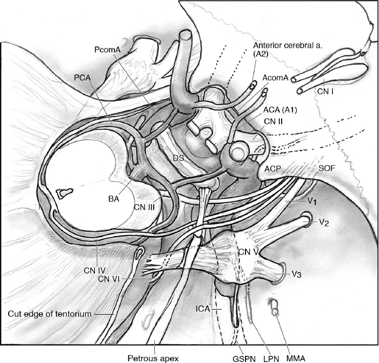
Fig. 2.6 Anatomic landmarks of the parasellar region. Compare the anatomic landmarks with the description of the skull base triangles in Table 2.4. ACA, anterior cerebral artery; AcomA, anterior communicating artery; ACP, anterior clinoid process; BA, basilar artery; CN, cranial nerve; DS, dorsum sellae; GSPN, greater superficial petrosal nerve; ICA, internal carotid artery; LPN, lesser petrosal nerve; MMA, middle meningeal artery; PcomA, posterior communicating artery; PCA, posterior cerebral artery; SOF, superior orbital fissure.
• Mastoid: a variably pneumatized bulbous bone (in the adult), the pneumatized cells of which are in direct communication with the middle ear via the aditus ad antrum (orifice of the mastoid antrum into the epitympanic recess).
 Koerner septum: Formed by the petrosquamous lamina, it is a thin bony septum that travels posteriorly from the epitympanum, separating the mastoid cavity into medial and lateral compartments.
 Muscular attachments:
 Sternocleidomastoid at the mastoid tip.
 Posterior belly of the digastric muscle at a sulcus just posterior to the stylomastoid foramen, often called the “digastric groove.”
• Tympanic: forms approximately three quarters of the bony external auditory canal in the adult (anterior wall, floor, and part of the posterior wall and roof).
 Surgical landmark: tympanomastoid suture line—the landmark for the exocranial facial nerve as the line curves inferiorly from the external auditory canal (EAC) in close proximity with the stylomastoid foramen.
 Anatomic landmark: petrotympanic suture—the chorda tympani exits from its intraosseous course through the suture/fissure.
• Petrous: forms the substrate of the middle and inner ear (Fig. 2.7) and has numerous surface landmarks as follows:
 Styloid process: a long bony process, 18 to 51 mm in length, that serves as an attachment point for muscles (stylohyoid, styloglossus, stylopharyngeus) and ligaments (stylohyoid, stylomandibular).22
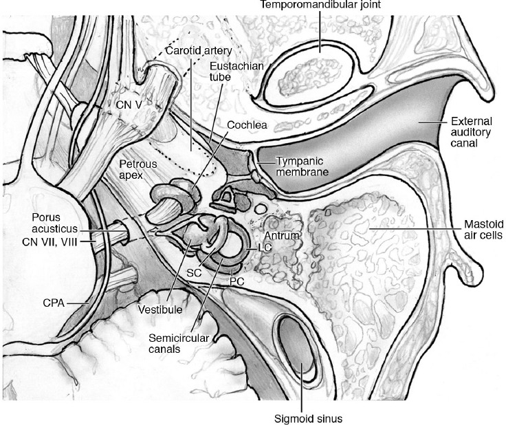
Fig. 2.7 Representation of the axial section of the petrous bone, showing the anatomic relationships with the middle fossa and cerebellopontine angle (CPA). Semicircular canals: SC, superior canal; PC, posterior canal; LC, lateral canal.
 Located anterior to the stylomastoid foramen (exit point of the facial nerve).
 Jugular fossa: Located medial to the styloid process and inferior to the middle ear cavity, the fossa is occupied by the internal jugular vein and nerves.
 Carotid canal: located directly anterior to the jugular fossa but is separated by a small wedge of bone. In the horizontal segment, the bone can be nonexistent on the superior surface.
 Sigmoid sulcus: The sulcus of the sigmoid sinus lies on the posterior intracranial surface and along the anterior limit of the posterior fossa.
 Arcuate eminence: Located superomedially on the intracranial surface, it signifies the superior semicircular canal, lateral to which is the roof of the middle ear and mastoid.
 Subarcuate fossa: situated superiorly and just posterior to the IAC and sometimes transmits the subarcuate artery.
 Depression of the trigeminal nerve (pars compacta): located on the superior surface of the petrous apex.
 Internal auditory canal (IAC): located on the medial aspect of the petrous portion. The medial opening is termed the porus acusticus internus, whereas the lateral end is termed the fundus.
 The fundus is divided into an upper and lower part by the horizontal crest (Fig. 2.8). The upper part is divided in two quadrants by a vertical bar (“Bill’s bar”). Considering four quadrants of the fundus (but anatomically the two lower ones are not separated by any bony structures), their contents are:
 Anterosuperior: facial and intermedius nerves
 Anteroinferior: cochlear division of CN VIII
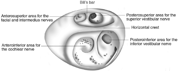
Fig. 2.8 Fundus of the internal auditory canal (IAC), right side. (Adapted from Koos WT, Matula C, Lang J. Color Atlas of Microneurosurgery of Acoustic Neurinomas. New York: Thieme; 2002.)
 Posterosuperior: superior vestibular division of CN VIII
 Posteroinferior: inferior vestibular division of CN VIII
 Groove of the endolymphatic duct: located posterolateral to the IAC within the posterior fossa and houses the endolymphatic duct and sac.
The Ear
• External ear: includes the auricle, the EAC, and the tympanic membrane.
 EAC: in adults, lateral one third is fibrocartilage; medial two thirds are bony, composed of the tympanic portion (anterior wall, floor, and part of the posterior wall and roof) and the mastoid portion of the temporal bone (remainder of the posterior wall and roof).
 Tympanic membrane (TM): typically measures 10 mm in size and is anchored to the EAC via the tympanic sulcus.
• Middle ear: an air-filled, mucous membrane–lined cavity containing the ossicles. The middle ear directly communicates with the mastoid cavity via the aditus ad antrum posteriorly, and the nasopharynx via the pharyngotympanic tube anteriorly.
 Boundaries:
 Anterior: pharyngotympanic tube (eustachian tube)
 Posterior: mastoid cavity
 Medial: otic capsule and promontory
 Lateral: tympanic membrane
 Superior: tegmen tympani (thin layer of bone separating the contents of the middle cranial fossa from the middle ear)
 Inferior: jugular wall (floor)
 Divisions of the middle ear23,24
 Epitympanum: also referred to as the attic and is the area superior to the level of the TM. The epitympanum communicates with the mastoid cavity via the aditus ad antrum
 Prussak’s space (superior recess): important region in acquired cholesteatoma.
 Subtended by the scutum and pars flaccida laterally, the neck of the malleus medially, and the lateral malleolar ligament superiorly.
 Mesotympanum: The area at the level of the TM and houses the majority of the ossicular chain.
 Ossicles: responsible for conductive hearing within the middle ear and amplify oscillations between the TM and the oval window.
 Malleus: composed of a head (articulates with incus), neck, anterior process, lateral process, and manubrium (attached to the TM).
 Incus: composed of a body (articulates with malleus), short process, long process, and lenticular process (articulates with stapes).
 Stapes: composed of head/capitellum (articulates with incus), an anterior and posterior crus, and a footplate (attached to the oval window).
 Suspensory ossicular ligaments: superior, lateral, and posterior malleal, and posterior incudal.
 Hypotympanum: area inferior to the level of the TM and contains the orifice of the pharyngotympanic tube (eustachian tube).
• Inner ear: consists of the bony labyrinth (otic capsule) and the membranous labyrinth that contains endolymph and is surrounded by perilymph. The bony labyrinth is very dense and represents the hardest bone in the body. The labyrinth consists of continuous subunits including the vestibule and three semicircular canals.
 Cochlea: Snail-shaped structure that tapers in width from base to apex during its 2½ to 2¾ turns; the basal, middle, and apical turns are separated by interscalar septa.
 Communicates with the middle ear at the oval window, which is abutted by the stapes footplate, and the round window membrane at the basal end of the cochlea
 Promontory: bulge of the basal turn of the cochlea that protrudes into the middle ear cavity that is visible on standard otoscopy through the TM.
 Jacobson’s nerve (branch of CN IX) runs over the middle ear surface of the promontory.
 Modiolus: highly porous crown-shaped bone at the core of the cochlea that enables passage of the auditory nerve fibers from the cochlear nerve to the organ of Corti.
 Cochlear aqueduct: a narrow bony passage that extends from the basal turn of the cochlea to the subarachnoid space in the posterior fossa that enables communication of perilymph and cerebrospinal fluid (CSF).
 Vestibule: ovoid-shaped structure that is situated posterior to the cochlea but is connected via the ductus reuniens.
 Within the vestibule are two end organs termed maculae, one oriented in the horizontal plane (utricle) and the other in the vertical plane (saccule).
 Relationships
 Anterior: cochlea
 Posterior: mastoid air cells
 Medial: posterior cranial fossa, into which the endolymphatic duct and sac extend
 Lateral: middle ear cavity (anterior) and mastoid air cells (posterior)
 Semicircular canals: three orthogonally oriented semicircular canals that project from the vestibule in a posterosuperior direction. Each semicircular canal has a dilated end, termed the ampulla, containing the receptors. The semicircular canals are the anterior (superior), posterior (dorsal), and horizontal (lateral).
 The anterior and posterior semicircular canals are vertically oriented and share a common crus, which opens into the superomedial part of the vestibule. The horizontal semicircular canal, however, has two separate openings into the vestibule.
Contents
• Musculature: Both the tensor tympani and stapedius muscles contract reflexively in response to loud noises to reduce excessive oscillation at the tympanic membrane and oval window, respectively.
 Stapedius
 Origin: within the pyramidal eminence (bony structure situated on posterior wall of middle ear)
 Insertion: head of the stapes
 Innervation: CN VII
 Tensor tympani
 Origin: within the superior aspect of the cartilaginous part of the eustachian tube
 Insertion: following a sharp turn at the terminus of the cochleariform process, inserts on the neck of the malleus
 Innervation: CN V3
• Neural Structures
 Sensory organs: anatomy described (see Inner Ear).
 Cochlea: anatomic structure responsible for the sensation of hearing via the organ of Corti, the primary sensory structure containing both inner and outer hair cells.
 Utricle and saccule: otolith organs within the vestibule responsible for the sensation of linear acceleration of the head in space.
 Semicircular canals: three orthogonally oriented structures responsible for vestibular function, specifically for the sense of angular acceleration/velocity (head rotation).
 Auditory and vestibular nerves (CN VIII): course through the IAC from the porus acusticus to the fundus and abuts the labyrinth.
 Cochlear nerve: traverses the modiolus to the cochlear end organ, the organ of Corti
 Superior vestibular nerve: supplies the anterior and horizontal semicircular canals and the utricle
 Inferior vestibular nerve: supplies the posterior semicircular canal and saccule
 Facial nerve (CN VII) (see Chapter 3, page 83).
 Greater superficial petrosal nerve: originates as the first branch of the facial nerve from the geniculate ganglion and carries presynaptic parasympathetic nerve fibers to the pterygopalatine fossa ganglion.
 Travels anteriorly and medially within the petrous portion of the temporal bone and emerges on its anterior surface via the hiatus for the greater petrosal nerve.
 Chorda tympani: conveys special sensory innervation to the anterior two thirds of the tongue and to the soft palate along with parasympathetic innervation to all salivary glands below the level of the oral fissure.
 Typically branches from the mastoid segment of the facial nerve just prior to its exit from the skull at the stylomastoid foramen.
 Ascends through the posterior canaliculus (canaliculus chordae tympani), which opens into the middle ear, and then arcs upward to cross the pars flaccida of the TM and passes between the neck of the malleus and the long process of the incus to reach the entrance of the anterior canaliculus (canal of Huguier or Civinini) above the insertion of the tensor tympani.
 The anterior canaliculus runs within the medial part of the petrotympanic fissure and exits into the infratemporal fossa.
 Nerve to stapedius: Innervates the stapedius muscle, as its name implies.
 Branches early from the mastoid segment of the facial nerve.
• Vascular
 External ear: arterial supply from the following branches of the ECA and venous drainage follows the arteries:
 Anterior auricular branches of the superficial temporal artery
 Posterior auricular artery
 Occipital artery
 Middle ear: primary arterial blood supply from the tympanic branch of the maxillary artery and the mastoid branch of either the posterior auricular or occipital arteries.
 Other arterial contributions from smaller branches of the following:
 Artery of the pterygoid canal
 Ascending pharyngeal artery
 Middle meningeal artery
 Tympanic branch of the ICA and rami caroticotympanici, whenever present
 Venous drainage into the pterygoid plexus and the superior and inferior petrosal sinuses.
 Inner ear: arterial supply is divided into vessels providing blood to the bony labyrinth and the membranous labyrinth, and venous drainage follows the arteries primarily into the inferior petrosal or sigmoid sinuses.
 Bony
Bony
 Anterior tympanic branch of the maxillary artery
 Stylomastoid branch of the posterior auricular artery
 Petrosal branch of the middle meningeal artery
 Membranous: Supplied by the labyrinthine artery which divides into:
 Cochlear branch
 Vestibular branch
 2.3 Topographic Anatomy of the Posterior Skull Base
Cerebellopontine Angle
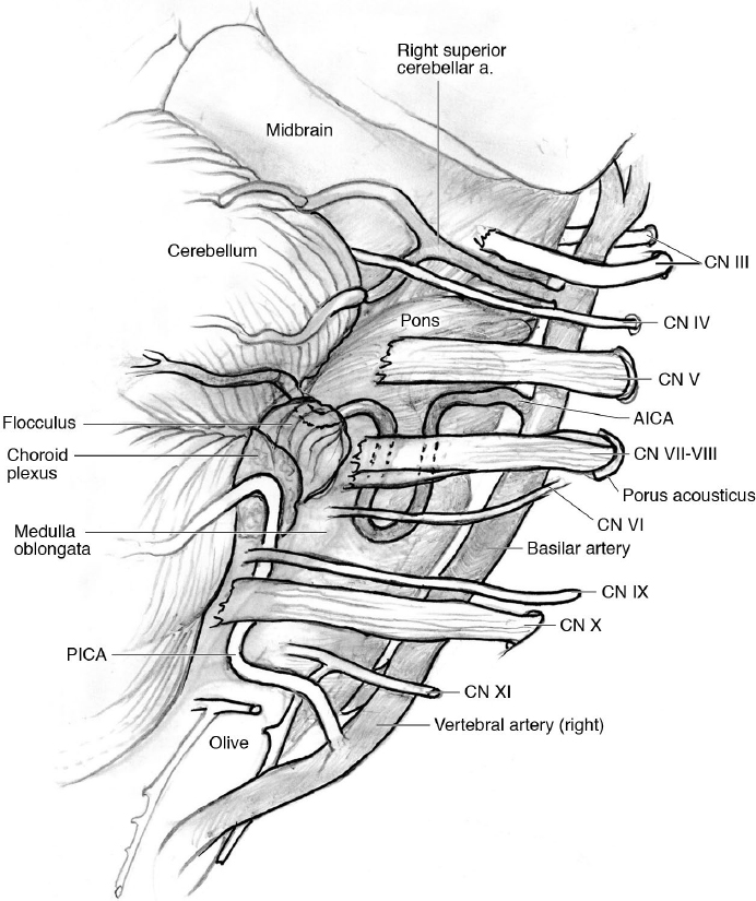
The cerebellopontine angle (CPA) is the anatomic space between the petrous bone and the petrosal cerebellar surface folding around the pons and middle cerebellar peduncle, containing the posterior cranial fossa nerves (Figs. 2.7 and 2.9).25 The structures in the CPA can be summarized as follows:
• Upper neurovascular complex: formed by the trochlear and trigeminal nerves, superior cerebellar artery (SCA) and its branches, and the petrosal veins complex.
 The motor root of the trigeminal nerve arises rostral to the sensory root. There are diffuse anastomoses between the sensory and motor component of the nerve, posteriorly to the ganglion of Gasser.
 The veins of the cerebellopontine fissure draining into the superior petrosal sinus are generally lateral to the trigeminal nerve.
 The superior petrosal veins drain into the superior petrosal sinus26,27: above and lateral to the IAC (type I), between the lateral limit of the trigeminal nerve and the medial limit of the facial nerve at the IAC (type II), and above and medial to Meckel’s cave (type III). Type II is the commonest arrangement, and type III is related to the best outcome in most retrosigmoid approaches that do not need to access regions medial to the entrance to Meckel’s cave.27
• Middle neurovascular complex: It includes the pons, middle cerebellar peduncle, cerebellopontine fissure, cerebellar petrosal surface, anterior inferior cerebellar artery (AICA), and CNs VI to VIII.
• Lower neurovascular complex: It includes the medulla, inferior cerebellar peduncle, cerebellomedullary fissure, vertebral artery and posterior inferior cerebellar artery (PICA), and glossopharyngeal, vagus, and spinal accessory nerves converging into the jugular foramen. The hypoglossal nerve is more medial and reaches the hypoglossal canal posteriorly to the vertebral artery.
Surgical Anatomy Pearl
All the inferior cranial nerves arise from rootlets exiting the brainstem in the post-olivary sulcus.
Foramen Magnum
The foramen magnum (FM) is oval-shaped and is delimited anteriorly by the clivus, laterally by the occipital condyles, and posteriorly by the anterior border of the occipital bone. The region of the FM includes the following:
• Cerebellar tonsils and inferior vermis
• Brainstem and rostral aspect of spinal cord
• CNs IX to XII
• Upper cervical nerves (C1 and C2)
• C1-C2 complex
• Vertebral arteries
• Posterior inferior cerebellar arteries
• Anterior and posterior spinal arteries
• Meningeal branches of the vertebral, external and internal carotid arteries
• Veins and dural sinuses of the craniovertebral junction
• Dentate ligaments
The hypoglossal canal is located above the occipital condyle.
Jugular Foramen
The jugular foramen is located on the floor of the posterior fossa, bordered anterolaterally and posteromedially, by the petrous bone and occipital bone, respectively. It is often morphometrically described as a triangular canal with exo- and endocranial openings that are located medial to the mastoid tip and tympanomastoid suture. The jugular spine or process splits the jugular foramen into the following two parts:
• Pars nervosa: smaller, anteromedial part that contains the CN IX and the inferior petrosal sinus.
• Pars venosa (pars vascularis): larger, posterolateral part that contains the internal jugular vein, jugular bulb, CNs X and XI, and the posterior meningeal branch of the ascending pharyngeal artery.
 2.4 Meningeal Folds
Anatomy of the Tentorium Cerebelli
The tentorium is a fold of dura mater forming the roof of the posterior fossa that separates the cerebellum from the cerebrum. Its free margin is the incisura, which is crossed by the brainstem and the cerebral peduncles. It slopes downward, like a tent, from its apex at the level of the posterior side of the incisura, to its attachment to the bones.
• Anterior borders: at the level of the petrous ridge on the posterior aspects of the petrous bone, where a tentorial division encloses the superior petrosal sinus.
The attachments of the tentorium on the petrous bone and clinoid processes form the anterior and posterior petroclinoid and interclinoid folds, resulting in the oculomotor triangle.28
 The falx cerebri fuses perpendicularly to the tentorium over its midline. The falcotentorial junction encloses the straight sinus, which receives venous blood from the vein of Galen and the inferior sagittal sinus. The straight sinus terminates posteriorly into the convergence of the sinuses at the level of the torcular herophili.
 The tentorial incisura has (a) anterior space, which is anterior to the brainstem, extending around the optic chiasm; (b) middle space, which is lateral to the brainstem, related to the hippocampal formation laterally; and (c) posterior space, which is posterior to the midbrain, in the region of the pineal gland and vein of Galen.28
 The average width of the incisura is about 30 mm, and the anteroposterior diameter is an average of 52 mm.
 The topographic neurovascular relationships of the tentorium and the tentorial incisura are with CNs III and IV, ICA, posterior cerebral artery (PCA), SCA, anterior choroidal artery, basal vein of Rosenthal, internal cerebral veins, vein of Galen, and straight sinus with the lateral and sagittal sinuses in the posterior part, at the level of the torcular.
Falx Cerebri (Cerebral Falx)
The falx cerebri is a thick fold of dura mater, attached anteriorly to the crista galli and posteriorly to the tentorium. Its superior margin is attached to the inner surface of the calvaria, and forms the superior sagittal sinus, whereas its inferior margin is free, containing the inferior sagittal sinus. The falx divides the two cerebral hemispheres.
Falx Cerebelli
The falx cerebelli is a dural fold below the tentorium cerebelli, projecting into the posterior cerebellar notch and into the vallecula of the cerebellum between the two cerebellar hemispheres.
Diaphragma Sellae
The diaphragma sellae is a horizontal meningeal fold attaching to the four clinoid processes, forming the roof of the sella turcica. It has an opening in the middle (ostium of the diaphragma sellae), crossed by the pituitary stalk (infundibulum). It separates the supradiaphragmatic space from the infradiaphragmatic space, where the pituitary gland lies.
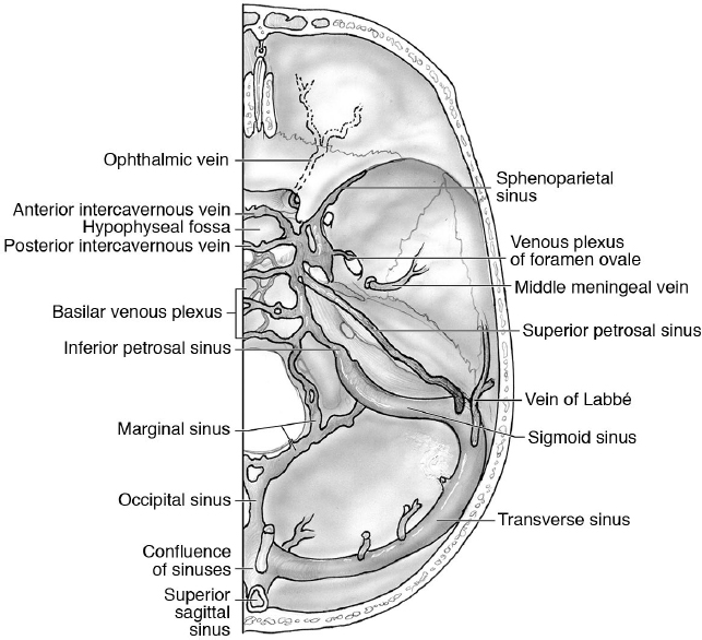
 2.5 Veins and Dural Venous Sinuses
See Fig. 2.10 and Chapter 26.
Transverse Sinus
The transverse sinus comprises symmetrical sinuses that drain blood from the superior sagittal sinus and from the straight sinus into the sigmoid sinuses. They begin at the internal occipital protuberance and, after a curvilinear course, reach the base of the petrous portion of the temporal bone.
Sigmoid Sinus
The sigmoid sinus is a paired sinus, beginning at the temporal bone, reaching the jugular foramen and draining into the internal jugular vein. It drains blood coming from the transverse sinus and petrosal sinuses, as well as from the vein of Labbé.