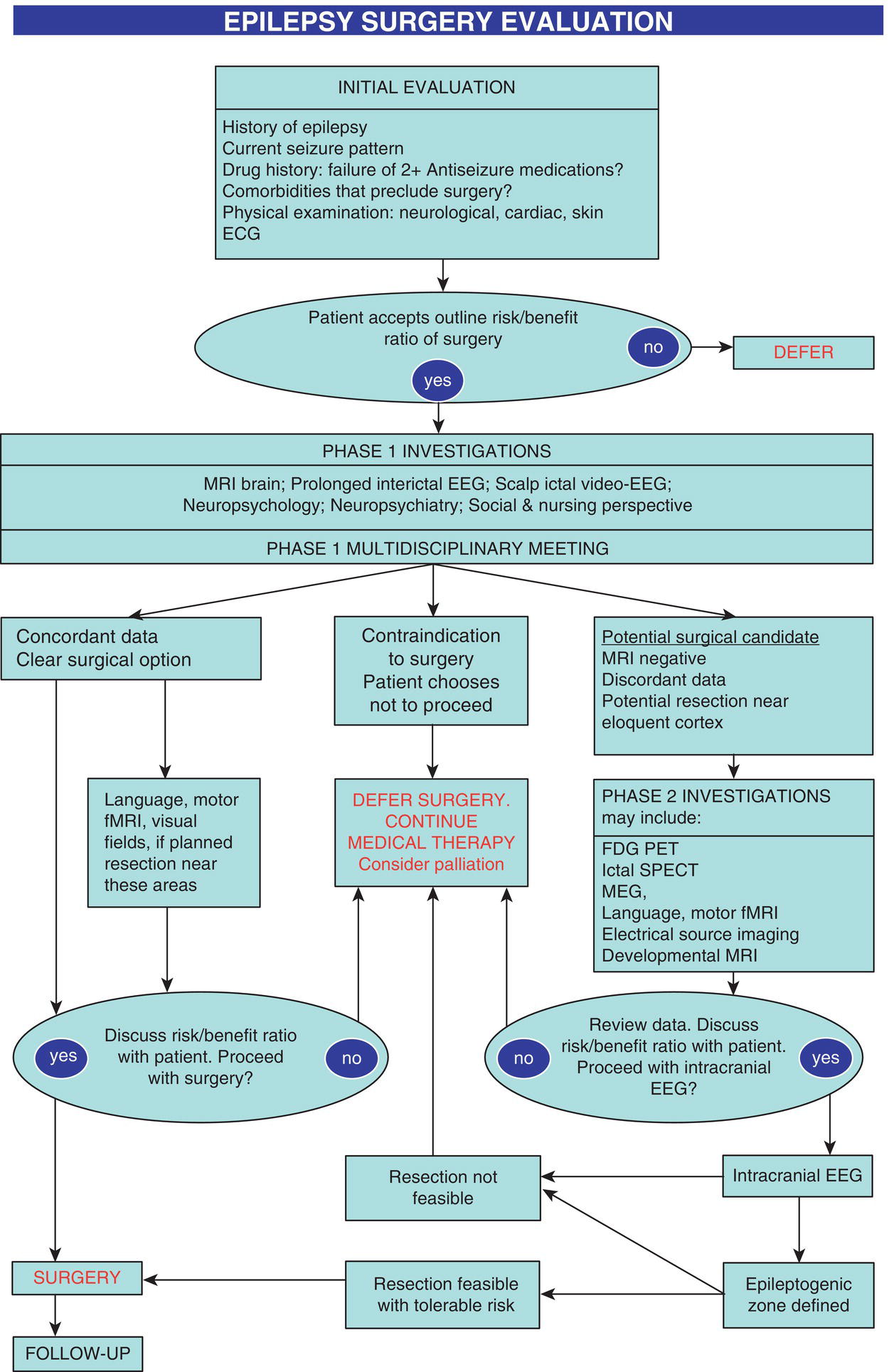
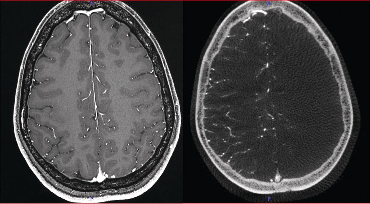
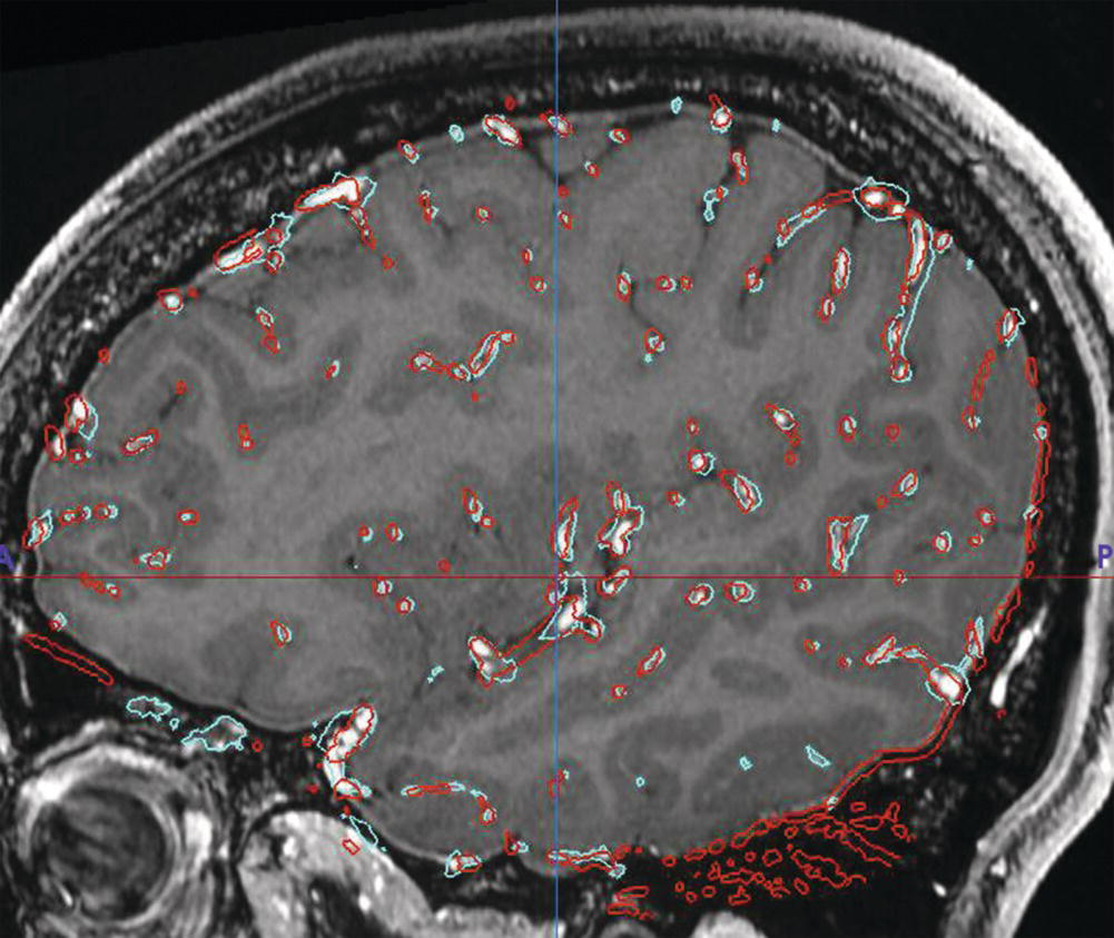
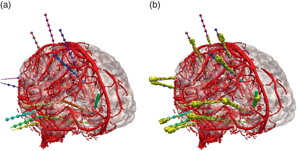
John S. Duncan, Anna Miserocchi, and Andrew McEvoy Surgery may be an effective treatment for drug‐resistant focal epilepsy (DRFE), but the current interval from epilepsy onset to surgery is an average of 18‐20 years.1 In a trial of surgery for temporal lobe epilepsy (TLE), two‐thirds became seizure‐free.2 In a study of TLE with a short duration, 85% of participants became seizure‐free.3 Prompt surgery provides the best opportunity for seizure remission, minimising adverse social and psychological consequences, and premature death. Machine‐learning routines have the potential to identify candidates for surgery earlier than is currently practised.4 The most common surgically tractable lesions underlying DRFE are hippocampal sclerosis (HS), focal cortical dysplasia, cavernoma, and dysembryoplastic neuro‐epithelial tumour. The best scenario is a discrete, safely resectable magnetic resonance imaging (MRI)‐visible lesion consistent with seizure semiology and the lateralisation and localisation of interictal and ictal EEG changes. The presurgical evaluation process (Figure 15.1) starts with an initial clinic visit, which involves reviewing the individual’s history, conducting a neurological and physical examination, and reviewing relevant clinical records. It’s crucial to provide the individual and their family with realistic expectations regarding the risks and benefits. Ideally, there is a 70% chance of remission, an additional 20% chance of significant improvement and a 1% risk of worsening seizures. Potential new neurological deficits include a 1% risk of substantial deficits, such as hemiparesis or dysphasia, and a 10% risk of lesser deficits, including a visual field defect that may preclude driving. There is also a risk of significant language or memory deterioration and a 50% risk of anxiety and depression in the months after surgery. If these potential outcomes are unacceptable, there is little merit in continuing, as investigation cannot improve these chances. Approximately half of people undergoing presurgical evaluation do not proceed to surgery. This is mainly because the epileptogenic zone (EZ), the smallest cortical part requiring resection for long‐lasting seizure remission, could not be defined, was multifocal, or resection would have led to an unacceptable deficit. Other reasons include a high risk of new morbidity and people choosing not to proceed.5 FIGURE 15.1 Surgery evaluation pathway. The ‘phase 1’ presurgical evaluation comprises the clinical history and examination, EEG, scalp video‐EEG telemetry, MRI, neuropsychological and neuropsychiatric assessments. Phase 1 results are reviewed at a multidisciplinary team meeting (MDT). At this stage, approximately a quarter of people may be directly recommended for resections. Half are advised not to proceed as it is impossible to localise a single EZ. Further ‘phase 2’ investigations are recommended in the remaining quarter to localise the EZ. These include developmental MRI acquisitions, post‐acquisition processing of MRI, PET, SPECT, electrical and magnetic source imaging and intracranial EEG. See Chapter 7 on neurophysiology and Chapter 8 on neuroimaging. Recording habitual seizures on video with scalp EEG to identify interictal epileptic discharges is crucial. Antiseizure medication (ASM) is commonly reduced by 50–75% to facilitate seizure occurrence. Reviewing the video with witnesses is critical to ensure that habitual episodes have been captured. A detailed analysis of seizure semiology, ictal and interictal EEG, is mandatory to infer the likely lateralisation and broad localisation of seizure onset. It is essential to keep in mind that, due to the cortex’s interconnectivity, seizure semiology may not provide a reliable or precise localisation of seizure onset, as seizures may start in a clinically silent area. See Chapter 3, The Spectrum of Seizures and Epilepsies, and Chapter 7, Neurophysiology. People with an identified epileptogenic lesion have 2.5 times higher odds of seizure freedom following surgery than if no lesion is identified. Advances in MRI, nuclear medicine and source localisation techniques help improve the delineation of the EZ. Surgically remediable lesions include hippocampal sclerosis (HS), developmental abnormalities, infections, neoplasia, stroke, trauma and vascular malformations. Optimised imaging acquisition and interpretation increase the detection of epileptogenic lesions. See Chapter 8, Neuroimaging. MRI acquisition protocols for epilepsy have been defined. The mainstay is high‐quality structural 3T MRI. See Chapter 8, Neuroimaging. Hippocampal sclerosis is the most commonly identified pathology in surgical series. It is characterised by hippocampal atrophy and increased T2 signal intensity. Visual inspection can miss subtle, focal or bilateral HS. T2 relaxometry quantifies the T2 relaxation time along the length of the hippocampus. 3D T2‐weighted FLAIR sequences may detect focal cortical dysplasia at the bottom of a sulcus, with blurring of the grey‐white boundary and dyslamination that extends into the white matter. Post‐acquisition MRI processing may enhance the detection of subtle abnormalities, but this increase in sensitivity comes at the expense of reduced specificity. If a detected lesion is concordant with clinical semiology, interictal and ictal video‐EEG, and satisfactory neuropsychological and neuropsychiatric assessments, no further investigation is usually required. If the planned resection margins are close to eloquent cortex, functional mapping, such as language and motor functional MRI (fMRI), and transcranial magnetic stimulation, may help delineate resection boundaries. Tractography can usefully demonstrate the relation to crucial tracts, including the corticospinal tract, optic radiation, arcuate, and inferior fronto‐occipital fasciculi. Commonly used fMRI language paradigms, verbal fluency, and verb generation primarily cause activation in the frontal lobe and lateralise language functions rather than precisely localising them. The sensitivity and specificity of fMRI for language lateralisation are 80‐90%. Picture naming and auditory naming primarily activate the temporal lobe and are better predictors of naming decline after anterior temporal lobe resection than are verbal fluency and verb generation.6 Anterior temporal lobe resections may cause visual field deficits that preclude up to half from driving even if they are seizure‐free. Tractography, derived from diffusion‐weighted MRI sequences, can visualise major white matter tracts such as the corticospinal tract and optic radiation. Embedding the tractographic representation of the optic radiation into the surgical microscope eyepiece during temporal lobe resection can prevent visual field defects from occurring.7 If MRI does not identify a lesion concordant with clinical and EEG data, functional imaging with 18F‐FDG PET and ictal‐SPECT may be helpful. Interictal PET has a sensitivity of up to 90% in TLE and 50% in extra‐TLE. The region of hypometabolism detected by 18F‐FDG PET is generally more extensive than the EZ and does not precisely outline a surgical resection plan.8 SPECT utilises technetium‐99m‐labelled ligands to measure regional cerebral blood flow (rCBF). The tracer can be administered at seizure onset and will distribute in the brain to reflect rCBF 40–60 seconds after the injection. Administering tracer as early as possible after seizure onset is crucial for identifying hyperperfusion associated with the seizure onset zone. Scalp EEG‐fMRI can detect haemodynamic changes associated with interictal epileptic discharges with a sensitivity of 30‐40% and may assist in planning intracranial implantation. The current clinical application of scalp EEG‐fMRI is the visualisation of interictal networks, which may help design intracranial EEG strategies and indicate a likely poor outcome, potentially justifying discontinuation of the evaluation. Magnetoencephalography (MEG) measures the magnetic field generated by synchronised post‐synaptic currents in pyramidal cell dendrites. In magnetic source imaging, current dipole maps of interictal spikes are overlaid onto an MRI scan. MEG’s spatial and temporal resolution is superior to scalp EEG but is limited to dipoles on the cortical surface and less sensitive to deeper sources. In people in whom the MEG signal was concordant with the resection, 85% were completely seizure‐free, compared to 37% when the MEG signal was not concordant. MEG has a limited role in helping to define the EZ, particularly in those with unremarkable MRI.9 The MDT meeting is an essential part of considering surgery, requiring active input from neurology, neurophysiology, neuroradiology, neuropsychology, neuropsychiatry, and neurosurgery. Careful documentation in a consistent format of evidence, options and rationale for recommendations is essential, particularly as multistage evaluations are complex and may extend over prolonged periods.10 Detailed and frank discussions with the individual are essential, and it is often advantageous to include the wider family. Nowhere is the principle ‘not about me, without me’ more appropriate than when weighing up the options for neurosurgical treatment and consideration of the risk–benefit ratio.11 Intracranial EEG recordings made directly from cerebral tissue are often required to localise the sites of seizure onset and spread, particularly when there is discordant imaging, clinical and scalp EEG data. This is usually necessary to define the extent of the EZ and the proximity of eloquent cortical functions, to design an optimal resection. Intracranial EEG recordings are mainly made using stereotactically placed depth electrodes (SEEG) or subdural grids and strips that record from the neocortical surface. SEEG enables sampling from deep and medial parts of the cerebral hemispheres. The morbidity of SEEG is about 1%. SEEG has a lower risk of complications and morbidity than do subdural grids.12 Planning SEEG electrode placement is time‐consuming and requires a multidisciplinary approach to ensure adequate sampling for testing a specific hypothesis. SEEG electrodes are meticulously planned to enter the brain on the crown of a gyrus, maximising distance from cerebral vasculature and avoiding sulcal pial boundaries. This strategic placement also aims for orthogonal skull entry, the shortest feasible intracranial length, and maximised grey matter contact, while maintaining at least a 10 mm separation from other implanted electrodes. Methods used to detect intracranial vasculature are varied, with digital subtraction angiography being the most sensitive but invasive. The visualisation of blood vessels with a diameter of 1 mm with MRI is now feasible (Figures 15.2 and 15.3). It has been recommended that there be a minimum of 3mm between blood vessels and electrode trajectories, particularly for vessels near the brain surface and those with a diameter greater than 1mm. The accuracy of SEEG implantation can be determined by co‐registering the preoperative plan with a post‐implantation CT scan, visualising the electrodes (Figure 15.4). Robotic guidance of SEEG placement achieved a median 0.8 mm entry point and 1.8 mm target point error, compared to a median 1.4 mm entry point and 2.7 mm target point error with a Talairach frame.13 Stereotactic surgical procedures depend on the accuracy and stability of individual registration to the device. A neuropsychological evaluation is essential in the presurgical assessment, providing information on laterality and localisation of cerebral function and dysfunction. A key role is to advise individuals about the cognitive impact of the proposed surgery. fMRI is used to lateralise language and memory functions. See Chapter 8, Neuroimaging. The tests used must be suitable for longitudinal reassessments to determine the impact of surgery on cognitive function. See Chapter 9, Neuropsychology. FIGURE 15.2 Left: Axial slice showing MRI vascular imaging T1 weighted with gadolinium. Right: Axial slice showing digital subtraction angiography from left internal carotid artery. FIGURE 15.3 Sagittal slice showing vascular 3D models derived from Digital Subtraction Angiography (red) and T1‐weighted MRI + Gadolinium (cyan) FIGURE 15.4 (a) Plan for SEEG into the right cerebral hemisphere. (b) the segmented electrodes, shown in yellow, after implantation. Inevitably, surgical resection and collateral damage from creating access to the resection target carry risks of cognitive impairment. More selective procedures have less chance of causing decline. Seizure freedom and reducing ASM, however, may enable cognitive improvement. Resecting highly functional cortex, as evidenced by high test scores and normal MRI structure, carries an increased risk of noticeable impairments. fMRI for episodic memory and naming is emerging as a predictor of verbal memory decline and word‐finding difficulty after anterior temporal lobe resection.14 After left temporal lobe resection, verbal memory is better preserved with smaller medial temporal resections, as the posterior hippocampus, inferior temporal and fusiform gyri have a crucial role in maintaining memory.15 Evidence of contralateral dysfunction and damage indicates limited cognitive reserve and reduced capacity for post‐operative improvement in function. Collateral damage caused by the open surgical approach has led to lesioning techniques, such as laser interstitial thermal therapy and thermocoagulation, which can cause less direct deficits by their approach. Preoperative psychiatric evaluation and post‐operative follow‐up are appropriate, considering the causes of psychopathology and treatment. Active psychiatric pathologies should be treated before surgical treatment as indicated. The main issues are 1. Is there a history of psychiatric illness that will reduce the chances of seizure freedom? 2. Does surgery increase the risk of psychiatric disorder? 3. Can surgery improve the psychiatric state? Interictal depression (5–50%), anxiety (0–48%), and interictal psychosis (0–16%) are common in individuals with DRFE.16 Structural brain damage, ongoing seizures, and adverse effects of ASM increase psychiatric morbidity, as do reactions to epilepsy and its psychosocial comorbidities. A history of psychopathology, particularly affective disorder, stress‐related disorders and substance abuse, is associated with a lower chance of seizure remission. Successful surgery may improve psychiatric symptoms, particularly postictal depression, psychosis and antisocial behaviour.17 Postoperatively, a transient increase in psychiatric symptoms, especially anxiety, is found in two‐thirds of individuals but is usually followed by long‐term improvement. The risk of post‐operative psychosis has been estimated at 3%. A presurgical psychiatric diagnosis is a risk factor for post‐operative psychiatric disturbance. De novo psychopathology has been noted in up to a quarter of people after TLE surgery (depression: 4–18%; anxiety: 3–26%; interictal psychosis: 1–12%).16 The incidence of de novo non‐epileptic seizures following surgery is estimated at 4%, higher in females with a psychiatric history. See Chapter 23, Neuropsychiatric Aspects.
CHAPTER 15
Epilepsy Surgery
EPILEPSY SURGERY EVALUATION
SCALP VIDEO‐EEG TELEMETRY
BRAIN IMAGING
MRI
FUNCTIONAL IMAGING
THE MULTIDISCIPLINARY EPILEPSY SURGERY TEAM
INTRACRANIAL EEG
NEUROPSYCHOLOGICAL EVALUATION
PSYCHIATRIC EVALUATION
Related posts:



Stay updated, free articles. Join our Telegram channel

Full access? Get Clinical Tree


