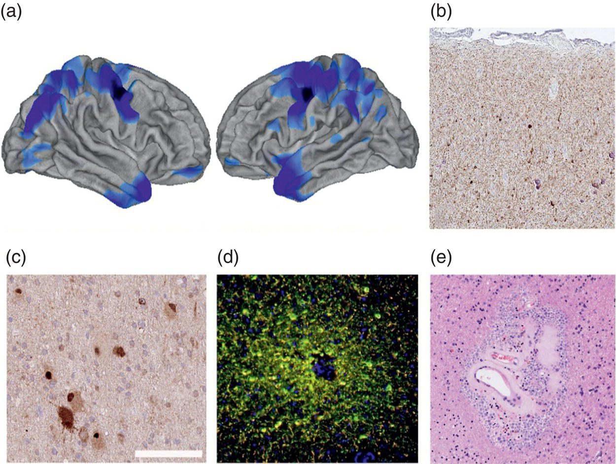
Maria Thom and Matthias J. Koepp Epilepsy is more common in older age. Alzheimer’s disease (AD) and epilepsy often co‐exist, and share significant mechanistic overlap. People with epilepsy are at a higher risk of dementia than the general population.1 Late‐onset epilepsy (LOE) is often accompanied by subclinical epileptiform activity, which can be a prodromal sign of AD.2 Seizures in people with AD may be subtle and misinterpreted as part of the disease process. Amyloid‐beta plaques, tau tangles, neuroinflammation, and mitochondrial dysfunction can alter neuronal activity, increasing seizure susceptibility. However, as a diagnosis of epilepsy is only made in up to half of people with AD, this could reflect underlying neuropathological differences, including regional variation in the disease stages. A recent study demonstrated that those with AD and more active seizures had greater brain tau and amyloid accumulation.3 Higher CSF tau and lower Aβ42, known AD severity markers, were noted in a large AD series with epilepsy compared to those individuals without seizures.4 If network hyperexcitability contributes to or exacerbates AD, increased tau and amyloid deposition should be anticipated in the epileptogenic hemisphere. A cross‐sectional study found a spatial association among the epileptogenic focus, tau, amyloid deposition, and neurodegeneration in early AD.5 People with temporal lobe epilepsy (TLE) often regard memory impairment as a more significant burden than seizures. Recurrent seizures in TLE may be associated with hippocampal atrophy, which can contribute to memory deficits. This damage, along with degeneration of the amygdala and entorhinal cortex, exacerbates cognitive impairments.6 TLE‐related neurodegeneration extends beyond the temporal lobes, affecting frontal and parietal cortices, which have a role in executive control and spatial memory. Antiseizure medications (ASM) may further exacerbate cognitive dysfunction, complicating diagnosis and treatment. Late‐onset epilepsy (LOE) increases the risk of AD and vice versa, but the precise temporal relationship between them remains unclear. Neuroimaging techniques, such as FDG‐PET, can help distinguish these conditions by identifying cerebral hypometabolism linked to brain areas shown on EEG to be generating seizures. Reduced glucose uptake was observed in the right posterior cingulate cortex and left precuneus areas in people with LOE compared to controls. This correlated with memory impairment, interpreted as indicative of AD‐related changes.7 PET imaging of neuroinflammation showed increased translocator protein binding in ipsilateral and contralateral hippocampi in TLE, possibly contributing to neurodegeneration.8 Neuroimaging studies have also investigated biomarkers such as P‐glycoprotein, amyloid‐beta, and tau. Longitudinal studies focusing on cognitive decline in epilepsy are needed.9 Amyloid tracers, such as PiB and flutemetamol, used for the diagnosis of AD, found higher amyloid in the prefrontal cortex of people with epilepsy and cognitive decline, particularly in APOE4 carriers, indicating its potential for diagnosing LOE‐related cognitive disease.10 CSF biomarkers are increasingly used in AD diagnostics, including Aβ and tau levels. Altered ratios of total CSF tau and phosphorylated tau (pTau) forms likely reflect tauopathy, while lower Aβ peptide levels likely reflect protein sequestered in plaques. CSF analysis in LOE, however, has shown inconsistent findings; some studies report pathologically low Aβ1‐42.11 and higher tau proteins7, while others report CSF levels similar to those of controls.12 As these studies are often cross‐sectional or at early time points, longitudinal studies of LOE and TLE cohorts with mapping of their cognitive trajectories are needed. Hippocampal atrophy is linked to memory impairments and cognitive decline.13 In addition to hippocampal damage, progressive cortical atrophy is a prominent finding, especially in drug‐resistant focal epilepsy (DRE).14 Neuroimaging studies suggest that neurodegeneration in focal DRE is progressive, with hippocampal, cortical, and subcortical changes being prominent (Figure 22.1a).15 Cortical thinning is not seen in unaffected relatives, suggesting this is an acquired phenomenon due to epilepsy‐related factors. Cross‐sectional studies linking the severity of brain atrophy to longer epilepsy duration indicate that damage accumulates over time.16 The ENIGMA–epilepsy consortium has shown common patterns of cortical atrophy in focal epilepsies.17 Longitudinal studies further confirmed that epilepsy leads to accelerated cortical atrophy. The thinning rate is more than double that of normal ageing, particularly in TLE and most pronounced in the first five years after epilepsy onset.14,18 Thinning was quicker in people over 55, suggesting age‐related vulnerability to epilepsy‐induced brain damage.18 These progressive cortical changes also occur in seizure‐free people, suggesting that neurodegeneration occurs independently of seizures, possibly driven by subclinical epileptiform activity. Epilepsy surgery, such as resection of the anterior temporal lobe, has been shown to slow the progression of cortical abnormalities, particularly in people who remain seizure‐free post‐surgery.19 Atrophy patterns in epilepsy differ from those seen in AD or chronic traumatic encephalopathy (CTE). Suggested mechanisms for this neurodegeneration include seizure‐related neuronal activity, accelerated ageing, minor traumatic head injuries, and degenerative vascular pathology, which may act synergistically. Functional neuroimaging has shown that longer epilepsy duration and higher seizure frequency are associated with reduced temporal lobe activation and impaired memory encoding.20 Magnetic resonance spectroscopy showed elevated glutamate and reduced N‐acetyl aspartate in atrophic regions, indicating neuronal dysfunction and cell loss.21 FIGURE 22.1 Neurodegeneration in epilepsy. (a) SPM analysis showing regions of significant cortical thinning compared to a control group on MRI in an adult cohort of 80 individuals with hippocampal sclerosis and temporal lobe epilepsy. Source: Courtesy of Fenglai Xiao. (b) pTau accumulation shown with AT8 antibody from an adult with Sturge Weber syndrome (leptomeningeal angiomatosis), showing FCD type IIIC in the adjacent perilesional cortex and regions of calcification in the surgically resected sample. (c) Evidence of defective autophagy pathways in balloon cells, as demonstrated by p62 immunohistochemistry, which reveals cytoplasmic and nuclear labelling in many atypical balloon cells and dysmorphic neurones in FCD type II. This condition also exhibits mTOR pathway activation. (e) Post‐mortem histology from a person with DRE and pTau accumulation around vessels in a pattern typical for chronic traumatic encephalopathy. This is demonstrated by multiplex Tau labelling, which shows the accumulation of three phospho‐tau types (AT8 in green, CP13 in yellow, and AT100 in red) that co‐localise in many neuritic processes. Source: Courtesy of Alicja Mrzyglod. (e) White matter of the temporal lobe from a person with TLE and hippocampal sclerosis. The white matter arterioles exhibited marked hyaline arteriolosclerosis and an abundant accumulation of corpora amylacea (also termed ‘wasteosomes’) in the perivascular spaces, which is considered a manifestation of impaired peri‐arteriolar drainage. Bar shown (in c): Equivalent to 500 microns in (b), 100 microns in (c), 200 microns in (d), 80 microns in (e). Neuroimaging provides crucial insights into neurodegenerative changes in epilepsy, including cortical atrophy and hippocampal damage. Neuroimaging techniques help to understand the pathophysiology of epilepsy and track disease progression, aiding in the development of interventions to mitigate brain damage and cognitive decline. The presence, type, severity, and trajectory of cognitive impairment in epilepsy vary significantly between individuals. This variation suggests varied pathological mechanisms, including seizure activity, underlying seizure causes, antiseizure treatments, seizure‐related trauma, vascular damage, activation of neuroinflammatory pathways, and the interplay of genetic factors. Specific syndromes, such as developmental and epileptic encephalopathies, progressive myoclonic epilepsies, and autoimmune encephalitis, are characterised by accelerating cognitive and neurological decline with distinct pathological brain alterations. Neurodegeneration, as a broad term, implies progressive neuronal and tissue loss, manifesting as atrophy. Extensive neuropathological studies in normal ageing populations highlight multimorbidity with vascular pathology and AD, which are frequently combined. Studies in epilepsy have mainly focused on samples from surgical resections. These capture degeneration at an earlier stage, exploring specific neuronal vulnerability, degenerative protein aggregation, glial and inflammatory responses, and vascular dysfunction, suggesting that multiple factors may contribute to memory decline. Neuronal loss is exemplified by hippocampal sclerosis (HS), characterised by a stereotypical pattern of subfield damage and gliosis. As the hippocampus plays a crucial role in memory encoding and is often included in surgical resections, it has been a focus of several studies. Studies correlating neuronal loss and HS patterns with memory decline have had conflicting findings.6 Reduced adult hippocampal neurogenesis in TLE has also been linked to cognitive verbal memory decline. The pathological correlates of progressive cortical thinning shown on MRI in epilepsy cohorts have been less explored and have a less clear impact on cognitive regression. A systems‐level analysis addressing cellular causes underlying patterns of regional cortical thinning in epilepsy identified activated microglia, suggesting neuroinflammatory pathways may contribute to the atrophy observed on MRI.22 There is controversy surrounding the role of tau in epilepsy. It is recognised that the peptides associated with AD, Tau and amyloid‐beta, can dysregulate neuronal and circuit hyper‐excitability. Tau oligomers and pathological forms enable epileptogenic activity in animal models, while tau reduction in excitatory neurons reduces seizures in Dravet models.23 Tau has many critical physiological functions, but its accumulation as misfolded and pTau aggregates is a hallmark of a disease state, known as a ‘tauopathy’. In the last decade, studies of tau in epilepsy surgical and post‐mortem tissues have shown: (1) overall lower ‘tau loads’ than in late‐stage AD;6,24 (2) age‐accelerated increases in tau in people with DRE compared to controls;25 (3) atypical distributions compared to Braak staging in AD; (4) less evidence for concomitant beta‐amyloid accumulation; (5) inconsistent association with cognitive impairment. The cause of any pTau accumulation in epilepsy may be multifactorial. In older people with TLE and HS, Tau may reflect coincidental age‐related tauopathies, such as primary age‐related tauopathy or AD. A recent proteomics comparison of hippocampal tissue from advanced AD and epilepsy showed similarities, with many of the altered proteins interacting with tau. This suggests that significant tau‐mediated effects in epilepsy could occur in the absence of cellular accumulation.26 Seizure‐induced tau accumulation, resulting from neuronal excitatory activity, could explain ‘non‐Braak’ neuronal vulnerability patterns, such as those observed in superficial cortical neurones, relative hippocampal sparing, and mossy fibre accumulation.24,26–28 Multifocal tau accumulation in superficial cortical neurones occurs in nodding syndrome, an endemic form of severe epilepsy found in East Africa.29 A superficial cortical pattern is also reminiscent of tau phosphorylation in late brain development, considered to enable microtubule plasticity during axonal growth. Localised phospho‐tau accumulation has also been reported with specific vascular lesions associated with seizures, including meningioangiomatosis (Figure 22.1b), which could implicate vascular dysfunction and hypoxia. Focal cortical dysplasia (FCD) type II, tuberous sclerosis lesions, and mTORpathies with activating mutations in mTOR pathway genes exhibit an early accumulation of phospho‐tau, particularly in abnormal cell types.30 mTOR activity indirectly regulates tau phosphorylation, degradation, and, importantly, its autophagic clearance. Recent single‐cell sequencing methods in FCD‐type II have identified a specific subset of dysmorphic neurones expressing genes associated with neurodegeneration.31 Accumulation of neuronal tau may also reflect a failure of cellular clearance. Impaired autophagy has been reported in FCD, characterised by excessive p62 expression in abnormal cell types, including balloon cells (Figure 22.1c), and the accumulation of lysosomal storage material.32 This was recently confirmed with spatial transcriptomics methods.33 Autopsies of people with epilepsy provide an opportunity to explore the extent and distribution of any neurodegenerative disease, including tau accumulation, after a lifetime of seizures. In an extensive post‐mortem study, age‐accelerated AD pathology was noted, highlighting a prior history of traumatic brain injury, resulting from ongoing seizures, as a risk factor.25 It also noted that pTau accumulation alone was not the only explanation for cognitive decline. In a large post‐mortem series of epilepsy cases, criteria for CTE neuropathology (CTE‐NC)34 were applied, showing 16% with perivascular, sulcal cortical tau accumulation and astroglial tau (Figure 22.1d).28 CTE‐NC correlated with evidence of head injury, which was presumed to be the result of poorly controlled seizures, with DRE a further risk factor. LOE was also associated with AD pathology in this series. There is uncertainty regarding the most pathogenic molecular forms of tau.35 Tau phosphorylation is considered a post‐translational modification relevant to its aggregation, with some phosphorylation sites enhancing this process. Tau types remain less explored in epilepsy than AD, but differential tau phosphorylation sites have been shown in experimental epilepsy models36 and TS, suggesting disease‐related signatures. Surgical epilepsy series to date have reported variable pTau types, broadly in line with those occurring in AD as well as physiological states (Figure 22.1d).6,24 Degenerative small vessel disease in white matter is a leading cause of dementia, including subcortical vascular dementia and also contributes to AD. Higher volumes of white matter hyperintensity, an MRI measure of vascular injury, are associated with increased LOE risk.37 They may contribute to epileptogenesis, including impaired cerebral perfusion and disruption of subcortical networks.38 White matter degeneration occurring in epilepsy may also contribute to cognitive deficits. Diffusion tensor imaging in epilepsy commonly shows abnormalities including increased mean diffusivity and reduced fractional anisotropy in white matter projection pathways, which are particularly pronounced in TLE with HS. Underlying pathological correlates of white matter changes include loss of myelin, axons, and oligodendrocytes, which have been linked to poor memory performance. A recent study of white matter diffusion alterations in TLE linked perivascular space enlargement to pre‐operative cognitive decline and to diffusion markers.39 Perivascular space enlargement may reflect dysfunction of the glymphatic system, which is responsible for clearing interstitial metabolites; failure of this system is also implicated in neurodegeneration. Small vessel arteriolosclerosis has been reported in epilepsy surgery series (Figure 22.1e).40 Altered vascular dynamics and blood–brain barrier insufficiency in experimental models and the progression of epilepsy in humans may represent a cause and a consequence of seizures. Blood–brain barrier impairment in seizures leads to degenerative changes in the vascular basement membrane, characterised by increased collagen and reduced aquaporin expression in astroglial cells.41 Pericytes, the perivascular contractile cells, undergo contractions and vasospasm in response to seizures,42 which could lead to intermittent alterations in local haemodynamics and tissue perfusion. Alterations of pericyte distribution have been observed in TLE and experimental models.39
CHAPTER 22
Epilepsy and Neurodegeneration
LATE‐ONSET EPILEPSIES
CSF BIOMARKERS
EVIDENCE OF NEURODEGENERATION IN MRI STUDIES
NEUROPATHOLOGY OF NEURODEGENERATION IN EPILEPSY
NEURONAL LOSS
TAU PATHOLOGY
Tau in epilepsy tissues
Coincidental tauopathy
Seizure‐related tau
Genetic factors
Traumatic brain injury during seizures and tau
Post‐translational modifications of tau in epilepsy
WHITE MATTER AND VASCULAR ALTERATIONS AS A CAUSE OF NEURODEGENERATION IN EPILEPSY
Microvascular structural alterations in epilepsy
Related posts:



Stay updated, free articles. Join our Telegram channel

Full access? Get Clinical Tree


