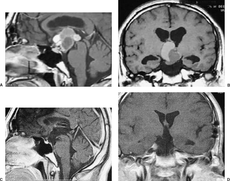
36 Diagnosis Large calcified craniopharyngioma Problems and Tactics Total resection of large craniopharyngioma with calcification is challenging. Large calcified portion has to be broken into pieces for resection. In the standard approach, such as interhemispheric or pterional approach, one cannot gain the space wide enough for insertion of tools such as a drill. The lesion was successfully removed with oticondylar transpersonal approach Keywords Craniopharyngioma, calcification, transpetrosal approach, middle fossa approach A 25-year-old man complained of headaches and nausea for a month prior to admission. Preoperative magnetic resonance imaging (MRI) revealed a calcified mass 2.5 cm in maximal diameter in the bottom of a large cystic tumor (5 × 4 × 3.5) in the third ventricle, extending up to the foramen of Monro (Fig. 36–1A,B). The patient was placed in a semiprone park bench position. A half generous coronal incision was made, starting from the inferior end of the ear lobe, running along the anterior margin of its cartilage and the parietal branch of the superficial temporal artery (STA) and vein (STV). An additional incision around the left ear lobe was then made (Fig. 36–2A). The pericranial flap pedicled with the STA and STV was dissected. The zygomatic arch, the lateral ligament of the temporomandibular joint, and the masseter fascia were exposed (Fig. 36–2B). The transected zygomatic arch hinged on the masseter was retracted downward. Following the lateral–suboccipital and temporal craniotomy, the outer layer of the mastoid process was split to keep the insertion of the sternocleidomastoid muscle on the lower part of the mastoid process (Fig. 36–2C) retracted downward. The oticocondylar osteotomy was done: condylar osteotomy consists of an osteotomy in the middle fossa 1 cm lateral to the foramen spinosum (Fig. 36–2D) and an additional osteotomy in the tympanosquamous suture (TS), and lateral to the tympanic membrane, crossing with the former osteotomy line (Fig. 36–2E). The temporal bone flap hinged on the pterigoid muscles was retracted downward (Fig. 36–2F). Then, following a presigmoidal approach1 (Fig. 36–2G), a durotomy was made along the posterior margin of the sigmoid sinus (SS) and the inferior margin of the superior petrosal sinus (SPS) leaving 5-mm dural fringes to these sinuses (Figs. 36–2G,H). A tentoriotomy was started along the posterior margin of the gassarian ganglion (GG) and directed to the dural entrance of the trochlear (fourth) nerve (N), dividing the SPS right anterior to the entrance of the petrosal vein (Fig. 36–2I). The arrachnoid membrane of the ventrolateral ambient cistern was opened to expose the left ventrolateral part of the infundibular portion between the posterior communicating artery (PCoA) and the oculomotor nerve (Fig. 36–3A, left and right). The third ventricular tumor was immediately encountered after midsagital incision of the very thin wall of the infundibulum, 10-mm in length. A hard, large, calcified mass was dissected, brought out over the pituitary stalk from the bottom of the tumor. During gentle holding of the calcified mass on the lateral side in its position by the tip of the suction tube, it was drilled by a diamond burr with a 3-mm round tip (Fig. 36–3B). The calcified mass shaved by the air drill was divided using a microscissors and the freed fragments were removed. The last sizable piece of the calcification, which was tightly adherent to the tumor with hard fibrous tissue, was separated and removed, using an ultrasonic aspirator. The remaining tumor was carefully separated, delivered by water dissection of the wall of the tumor capsule2 from the adherent third ventricular wall using a simultaneous suction and irrigation device (Opt irrigator, produced by Muranaka Co., Osaka, Japan) (Fig. 36–3C, left and right).
Oticocondylar Transpetrosal Approach (an Extensive Middle Fossa Approach) for Radical Removal of a Large Calcified Craniopharyngioma
Clinical Presentation
Surgical Technique
Related posts:
 28 Dominant Hemisphere Insular Tumor
 9 Large Aneurysm of the Internal Carotid Artery Obliterated with Seven Fenestrated Clips under Intraoperative Monitoring of Anterior Choroidal Arterial Blood Flow Insufficiency
 20 Collateralization via Vasa Vasorum: A Determinant of Therapeutic Efficacy of Coil Embolization of the Thrombosed Giant Aneurysm of the Vertebral Artery
 19 Partially Thrombosed Giant Dissecting Aneurysm of the Vertebral Artery—Treatment Strategies
 18 Combined Endovascular and Surgical Treatment of a Large Basilar Trunk Aneurysm in a Child
 70 Fibrous Dysplasia of the Paranasal Sinuses and Anterior Cranial Base
Stay updated, free articles. Join our Telegram channel

Full access? Get Clinical Tree


