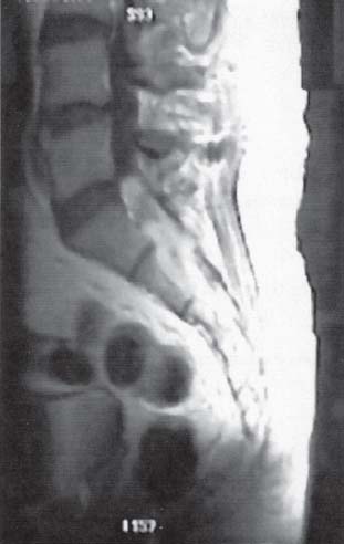
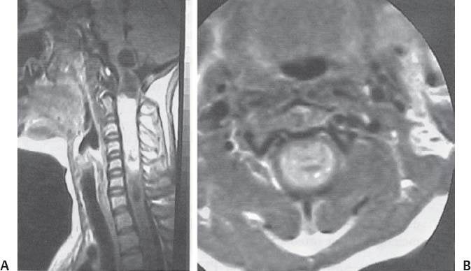
C H A P T E R 4 SPINAL TUMORS I. PRIMARY SPINAL TUMORS A. Ependymoma—the most frequent tumor in the lower spinal cord 1. Treatment—surgical resection; use intraoperative nerve root monitoring (avoid paralytics) 2. Lesions of the cauda equina—section the upper filum first to prevent retraction (Fig. 4.1) Fig. 4.1 Myxopapillary ependyoma. Sagittal-Infused, T1-weighted magnetic resonance image demonstrates enhancing nodular mass filling the distal spinal cord. (With permission from Citow JS. Neurosurgery Oral Board Review. 1st ed. New York, NY: Thieme Medical Publishers; 2003: 74, Fig. 9.5.) Fig. 4.2 (A,B) Intramedullary juvenile pilocytic astrocytoma. (A) Sagittal- and (B) axial-infused, T1-weighted magnetic resonance images demonstrate the enhancing tumor with the associated syrinx extending into the brainstem. (With permission from Citow JS. Neuropathology and Neuroradiology: A Review. New York, NY: Thieme Medical Publishers; 2001: 195, Fig. 268.) B. Astrocytoma—more common in children and in the upper spinal cord; usually low grade 1. Treatment—surgery (using somatosensory evoked potential [SSEP] monitoring) and radiation (XRT; only if not low grade) (Fig. 4.2A,B) C. Hemangioblastoma—usually cystic with a vascular mural nodule and located near the cord surface 1. Treatment—surgical resection D. Vertebral hemangioma 1. Treatment (only if symptomatic)—XRT, embolization, vertebroplasty, and rarely surgical resection E. Multiple myeloma (an isolated lesion is called a plasmacytoma) 1. Evaluation—urinalysis (Bence Jones protein), complete blood count (CBC; anemia), blood serum electrophoresis (IgG kappa chains), and skeletal bone survey 2. Treatment—XRT and chemotherapy F. Metastatic tumors 1. Treatment a. Surgery—indicated for unknown diagnosis, decompression, stabilization, and recurrence despite radiation b. Steroids and XRT—30 Gy over 10 days, two levels above and below the tumor Helpful Hints
4: SPINAL TUMORS
Only gold members can continue reading. Log In or Register to continue


Full access? Get Clinical Tree


