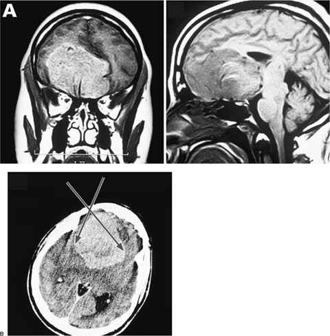
43 Diagnosis Frontobasal meningioma Problems and Tactics A huge frontobasal tumor was diagnosed in a 34-year-old man. After careful preoperative planning taking into consideration the individual pathoanatomical situation, the tumor was exposed via right supraorbital craniotomy. The supraorbital approach allowed wide intracranial exposure of the frontobasal tumor, according to the concept of keyhole approaches. The limited craniotomy offered minimal brain retraction thus significantly decreasing approach-related morbidity. In addition, the short skin incision within the eyebrow and the careful soft tissue dissection resulted in a pleasing cosmetic result for the patient. Keywords Frontobasal meningioma, supraorbital craniotomy, surgical approach This 34-year-old man was admitted in a neurological department with headaches, progressive disturbance of memory, apathy, inactivity, and aggressiveness. The neurological examination revealed slight organic brain syndrome and right-sided anosmia. Cranial magnetic resonance imaging (MRI) demonstrated a huge bifrontal frontobasal tumor extending mostly to the right side, with an enormous space-occupying effect (Fig. 43–1A). Given the progressive psychological syndromes and space-occupying effect, urgent surgical removal was indicated. In general, in cases of large intracranial tumors, the optimal surgical corridor corresponds to the length axis of the lesion. Using the sectorlike widening of the visual field, these extended, especially deep-seated lesions can be safely approached through small, well-placed craniotomies (Fig. 43–1B). In this illustrative case, the right-sided, frontobasal meningioma with a longitudinal tumor axis was exposed via a limited right supraorbital craniotomy. Along the frontal skull base, the tumor matrix was coagulated and resected step by step, and the meningioma was removed piecemeal. After induction of endotracheal anesthesia, the patient was placed in the supine position with the head elevated ~15 degrees. Thereafter, the head was rotated 45 degrees to the left and retroflected ~20 degrees. This head position allowed an ergonomic working position during surgery; the maneuver of retroflexion supported gravity-related self-retraction of the frontal lobe. After fixation of the head with a special radiolucent three-pin Mayfield holder, computed tomography (CT) was performed using mobile CT (Philips Tomoscan M, The Netherlands) dedicated for intraoperative application (Fig. 43–1B). The portable gantry was used in the later course of the operation, controlling the radicality of tumor removal. FIGURE 43–1 (A) Pre-operative magnetic resonance images (MRIs) in the coronal and sagittal planes of a 34-year-old man, clearly showing the enormous space-occupying effect of the solid, homogeneous, encapsulated, frontobasal meningioma, which extends mostly to the right. (B) Intraoperative computed tomographic scan showing positioning of the head before craniotomy. According to the longitudinal tumor axis, the sector-like surgical dissection allowed a safe approach to the large meningioma. Note the extension of the limited supra-orbital craniotomy (arrows).
Supraorbital Keyhole Craniotomy for Frontobasal Meningiomas
Clinical Presentation
Surgical Technique
Stay updated, free articles. Join our Telegram channel

Full access? Get Clinical Tree


