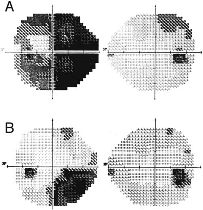
46 Diagnosis A clinoidal meningioma Problems and Tactics A 4-cm meningioma was found at the left anterior clinoid process in a 38-year-old woman who complained of visual impairment in the left eye. The tumor was demonstrated to extend into the left optic canal. Complete tumor removal and preservation of vision were attempted. Keywords Anterior clinoid process, meningioma, optic nerve This 38-year-old woman had complained of visual deterioration in the left eye. Her vision of the right eye had been mildly poor since childhood. Neuroophthalmo-logic examination revealed that the vision was 0.7/0.9 and the visual field of the left eye was defected on the nasal side (Fig. 46–1A). Magnetic resonance imaging (MRI) showed a well-enhanced mass at the left anterior clinoid process, and the maximum diameter was 41 mm (Fig. 46–2A). The obliquely sagittal plane showed the tumor penetrated into the left optic canal (Fig. 46–2A). The patient was placed in the supine position. A left frontotemporal craniotomy was performed. Bridging veins between the temporal lobe and sphenoid ridge were isolated and freed from their surroundings to enable multidirectional surgical access to the tumor by mobilizing the temporal lobe. The tumor was soft and strongly adherent to the base of the frontal lobe. The tumor was partially resected and a left optic nerve was found displaced medially. Removal of the anterior clinoid and optic unroofing were performed with a “protective dural flap” method (described following here) for removing the tumor in the optic canal and the paraclinoid tumor attachment. A semicircular dural flap was made by cutting the dura along the tumor margin and was pulled with two threads over the anterior clinoid process and optic canal (Fig. 46–3). The flap was extended over the underlying optic nerve and tumor with tapered spatulas to create a space for drilling the bone. The site for drilling was adequately exposed and bone was drilled away smoothly without compressing the optic nerve. The dural flap was divided between the left optic nerve and the internal carotid artery (ICA) by pulling the threads separately, and the tumor was found in the lateral side of the optic canal. After removing the tumor in the canal and around the proximal ICA, the tumor attachment was coagulated and removed. The anterior cerebral artery (ACA) was found encased by the tumor. The tumor was dissected from the ICA, posterior communicating artery (PCoA), middle cerebral artery (MCA), and ACA. Several perforators from the PCoA and ACA were sharply dissected from the tumor with microscissors. The tumor was found adherent to the undersurface of the left optic nerve and the anterior side of the pituitary stalk. The tumor was removed in piecemeal fashion from those critical structures, with the arachnoid membrane preserved as much as possible. Vascular and neural structures were not damaged by the procedure. After confirmation of complete hemostasis, the wound was closed in layers. FIGURE 46–1 (A)
Visual Improvement in a Clinoidal Meningioma Extending into the Optic Canal
Clinical Presentation
Surgical Technique
Related posts:
 28 Dominant Hemisphere Insular Tumor
 9 Large Aneurysm of the Internal Carotid Artery Obliterated with Seven Fenestrated Clips under Intraoperative Monitoring of Anterior Choroidal Arterial Blood Flow Insufficiency
 20 Collateralization via Vasa Vasorum: A Determinant of Therapeutic Efficacy of Coil Embolization of the Thrombosed Giant Aneurysm of the Vertebral Artery
 19 Partially Thrombosed Giant Dissecting Aneurysm of the Vertebral Artery—Treatment Strategies
 18 Combined Endovascular and Surgical Treatment of a Large Basilar Trunk Aneurysm in a Child
 70 Fibrous Dysplasia of the Paranasal Sinuses and Anterior Cranial Base
![]()
Stay updated, free articles. Join our Telegram channel

Full access? Get Clinical Tree


