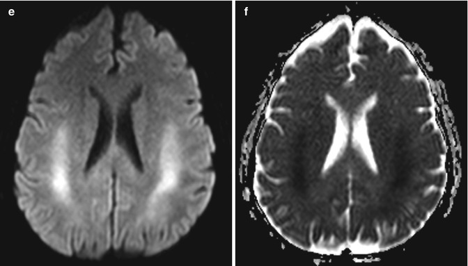
Fig. 20.1
5-FU-induced leukoencephalopathy. The patient presented with acute mental status changes during the course of therapy. Axial (a–c) and coronal (d) FLAIR MR images show extensive bilateral symmetric confluent high T2 signal involving the periventricular and deep cerebral white matter, including the corpus callosum, as well as the cerebellar white matter. DWI (e) and ADC map (f) obtained a few days earlier show areas of restricted diffusion in the cerebral white matter, which preceded other abnormalities. Treatment was discontinued and the abnormalities resolved on follow-up imaging (not shown)
20.4 Differential Diagnosis
Toxic leukoencephalopathy can be caused by other chemotherapeutic agents, such as methotrexate (refer to Chap. 19), vincristine, ifosfamide, fludarabine, cytarabine, cisplatin, and the interferons. This condition may also resemble the leukoencephalopathy associated with other pharmaceuticals, such as methotrexate (refer to Chap. 19), metronidazole (refer to Chap. 25) and vigabtrin (refer to Chap. 29), as well as various illicit drugs, such as opioids and amphetamines (refer to Chaps. 6 and 7). Otherwise, 5-FU toxic leukoencephalopathy may resemble other demyelinating conditions, such as acute disseminated encephalomyelitis caused, for example, by vaccines (refer to Chap. 48) and hypoxic-ischemic encephalopathy (refer to Chaps. 3, 9, and 40).
Related posts:


Stay updated, free articles. Join our Telegram channel

Full access? Get Clinical Tree


