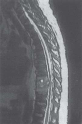
C H A P T E R 5 OTHER SPINAL DISORDERS I. SPINAL ARTERIOVENOUS MALFORMATIONS (AVMS) A. Type I—dural artery to a spinal vein in a foramina 1. Most frequent type (70%) 2. Usually occurs in older men 3. Causes progressive myelopathy 4. Foix-Alajouanine syndrome—subacute necrotizing myelitis due to venous hypertension B. Type II—glomus AVM is intramedullary. C. Type III—juvenile AVM is intra- and extramedullary. 1. Carries a worse prognosis D. Type IV—intradural perimedullary arteriovenous fistula (AVF) 1. Occurs in younger patients 2. Frequently hemorrhages E. Evaluation—magnetic resonance imaging (MRI) and angiography 1. Type I and IV AVMs—usually in adults; present with progressive weakness by venous hypertension 2. Type II and III AVMs—usually in children F. Treatment—endovascular embolization of feeding vessel or open coagulation and division of feeding vessels 1. Type I AVMs—radicular artery can be divided or clipped on the foramen. II. SYRINGOMYELIA A. Communicating—central canal is patent (i.e., Chiari malformation). B. Noncommunicating—trauma or tumor C. Signs/Symptoms—capelike, suspended, dissociated sensory loss (of pain and temperature); pain; Charcot joint; upper motor neuron findings in the lower extremities and lower motor neuron findings in the upper extremities D. Evaluation—MRI and possibly cine-MRI E. Treatment—occipitocervical decompression with duraplasty in the presence of a Chiari malformation (over 80% of these syrinxes will resolve without shunting) 1. Other types of syrinxes—treat by local decompression if possible or syringoperitoneal shunting (midline or dorsal root entry zone incision with 26-gauge tubing) 2. Syringosubarachnoid shunting—less successful III. PAGET DISEASE A. Cause—increased resorption of bone and replacement with irregular weak bone B. Signs/Symptoms—bone pain, fractures, back pain, and deafness 1. Occurs in the skull, axial, and long bones 2. Rarely develops into a sarcoma C. Evaluation—increased serum alkaline phosphatase, normal calcium, increased urine hydroxyproline from cartilage, bone scan, and x-ray (thickened bones and lytic skull) D. Treatment—calcitonin, bisphosphonates, and surgical decompression (there is frequently much blood loss) A. Cause—usually S. aureus. Risk increased in the obese, elderly, ill, diabetic, or immunosuppressed B. Treatment 1. Culture-guided antibiotics—14 days for superficial infection 2. Surgical debridement 3. Closing over a drain or packing 4. Plastic surgery closure—consider for infection extending down to the bone V. SPINAL EPIDURAL ABSCESS A. Causes—usually due to staph rather than strep; most often seen in intravenous drug abusers and diabetics B. Signs/Symptoms—pain, tenderness, fever, and focal symptoms related to pressure or thrombophlebitis C. Evaluation—MRI, complete blood cell count (CBC), and erythrocyte sedimentation rate (ESR) D. Treatment—antibiotics, immobilization, and decompression VI. VERTEBRAL OSTEOMYELITIS A. Causes—usually staph, strep, or TB B. Treatment—antibiotics and immobilization (Fig. 5.1) VII. DISKITIS A. Causes—staph and strep B. Treatment—antibiotics and immobilization Fig. 5.1 Osteomyelitis. T2-weighted magnetic resonance image of the thoracic spine demonstrating Pott disease with diskitis and osteomyelitis at T7 and 8. (With permission from Citow JS. Neuropathology and Neuroradiology: A Review. New York, NY: Thieme Medical Publishers; 2001: 199, Fig. 272d.) Helpful Hint
5: OTHER SPINAL DISORDERS
Only gold members can continue reading. Log In or Register to continue


Full access? Get Clinical Tree


