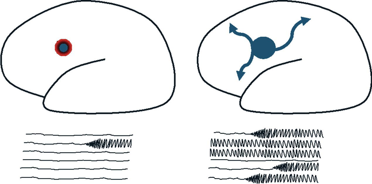
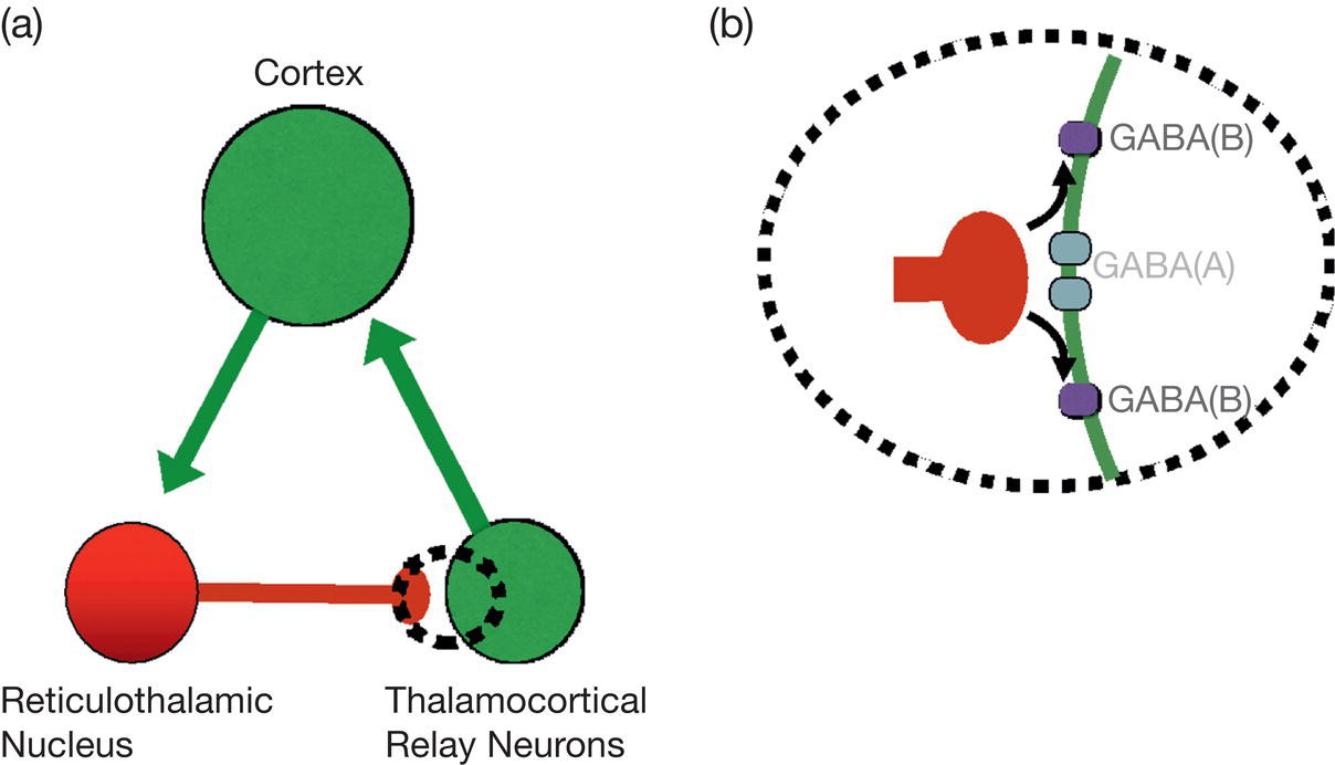
Matthew C. Walker and Dimitri M. Kullmann Despite our burgeoning understanding of epilepsy mechanisms, John Hughlings Jackson’s 150‐year‐old definition of epilepsy broadly holds to this day: ‘Epilepsy is the name for occasional, sudden, excessive, rapid, and local discharge of grey matter’. The epilepsies are considered a group of conditions broadly divided into focal and generalised epilepsies, and seizures are now defined as ‘a transient occurrence of signs and or symptoms due to abnormal, excessive or synchronous neuronal activity of the brain’. Seizures are distinct entities characterised by specific defining features. The discharge described by Hughlings Jackson often does not remain ‘local’ as exemplified by his eponymous Jacksonian march, with the recruitment of contiguous areas in the primary motor cortex. Focal seizures often evolve, as different (and sometimes distant) brain areas are recruited, leading, in some cases, to a bilateral tonic–clonic seizure. Seizures are also marked by their brevity, usually lasting seconds or minutes, suggesting that there are processes which actively terminate seizure activity; failure of these processes leads to status epilepticus. It is also important to distinguish seizures from epilepsy, which is the propensity to have seizures. These considerations call for a mechanistic understanding of several processes. These include the initiation, spread and termination of seizures, and epileptogenesis, which describes the transition from a non‐epileptic brain to one that can generate seizures. Epilepsy itself can also worsen with time; a phenomenon intimately related to epileptogenesis. We consider the mechanisms by which focal seizures arise in the cortex and escape inhibitory restraint, as well as the mechanisms underlying thalamocortical seizures, which affect both hemispheres simultaneously. We then consider the processes underlying epileptogenesis. In all these studies, much of the work has been associational and establishing causal relationships remains challenging. Even when a genetic mutation is at the root of the epileptic disorder, precisely how and where in the brain this leads to seizures is often difficult to determine. The normal functioning of neuronal circuits depends on an intricate interplay between excitatory and inhibitory mechanisms. Experimental manipulations interfering with inhibition are a powerful trigger for seizures, and conversely, many antiseizure medications (ASM), such as benzodiazepines and barbiturates, act by promoting inhibition. Similarly, manipulations that enhance neuronal or synaptic excitation are proconvulsant. The most common mechanism of action of ASM is to reduce neuronal excitability, for instance by promoting the inactivation of sodium channels (e.g. phenytoin, carbamazepine, lamotrigine, lacosamide), and perampanel directly reduces glutamatergic synaptic transmission.1 Epilepsy is often described as a state of ‘excitation/inhibition imbalance,’ but this concept does not capture the full complexity of instability characterising the disorder. First, inhibition cannot be equated with the primary inhibitory neurotransmitter, GABA, or GABAergic interneurons. Other neuroactive molecules, such as adenosine2 and some neuropeptides3 exert powerful inhibitory effects on principal excitatory neurons. GABAergic interneurons do not exclusively innervate principal neurons. A subset preferentially targets other interneurons, and there are instances in which GABAergic transmission can be excitatory; moreover, GABAergic inhibition synchronises the activity of networks of excitatory neurons.4 Inhibition can also be considered at the level of individual neurons. Potassium channels, for instance, generally act to maintain the resting membrane potential of neurons, countering the effects of excitation and inhibiting the generation of action potentials.5 4‐aminopyridine (fampridine), a blocker of potassium channels, is a powerful chemoconvulsant when applied directly to the brain. ASM that activate potassium channels are in clinical trial6 These mainly target the same potassium channel subtype as retigabine, which was withdrawn because of side effects. The concept of ‘excitation/inhibition imbalance’ alone does not explain why seizures are generally brief and intermittent phenomena in most people with epilepsy. Even in those with daily seizures, the brain spends over 99.9% of the time not generating seizures. The concept of ‘inhibitory restraint’ explains how local over‐excitation is contained by inhibitory mechanisms, which can intermittently fail (Figure 6.1), Epilepsy is better considered as an ‘instability’ rather than as an ‘imbalance’.7 Principal neurons may be overexcited because of various pathological processes such as disrupted neuronal migration in focal cortical dysplasia, aberrant axonal and synaptic connections, or loss of a subset of inhibitory interneurons. Rare causes such as autoantibodies or mutations affecting LGI1 may also predispose to over‐excitation.8 Regardless of the underlying abnormality, this may trigger a positive feedback loop, giving rise to excessive and persistent recruitment of principal neurons. At the cellular level, a sudden paroxysmal depolarising shift of principal neurons is observed, maintained by excitatory synaptic activity and the intrinsic excitability of neurons.9 Overactivity of principal neurons is usually contained by the rapid recruitment of GABAergic interneurons in the immediate vicinity, which is analogous to a fire blanket.10 It is speculated that such events correspond to interictal epileptiform discharges, typically lasting less than 200 ms, which are a frequent biomarker of epileptogenic tissue. Experimental data indicate that such spikes are associated with GABA release, likely originating from an annulus of interneurons that are brought to the firing threshold by the synchronous or near‐synchronous firing of a population of principal neurons. The effectiveness of this inhibitory recruitment has led to such discharges being considered ‘inhibitory,’ and interictal epileptiform discharges may serve as an indicator of effective inhibitory restraint. FIGURE 6.1 Inhibitory restraint. Initiation and propagation of seizures are prevented by an inhibitory restraint mainly due to the activation of GABAergic inhibitory neurons, so that excitation (blue) is constrained by an annulus of inhibition (red) (left panel). If this inhibition breaks down, seizure activity can then propagate to other regions (right panel). Since there are such effective and robust inhibitory mechanisms, why do seizures occur, and how do they spread? Evidence indicates that seizure initiation and spread may have multifarious and distinct mechanisms. These mechanisms may vary between brain regions and different types of epilepsy. In any brain, the excessive recruitment of principal cells will eventually lead to escape from inhibitory restraint (Figure 6.1).11 An example is electroconvulsive therapy in which current is passed through the brain, resulting in excessive and synchronised neuronal recruitment, leading to a generalised convulsion. Two processes may be seizure‐promoting: the excessive activation of principal neurons and the failure of GABAergic inhibition. In some models, observations of seizure initiation suggest that intense GABAergic recruitment precedes excessive principal cell recruitment. What processes underpin the failure or pro‐seizure effect of GABAergic inhibition? Several mechanisms have been proposed and may contribute variably to the generation of different seizures with different aetiologies. These mechanisms (failure of GABA release, a net pro‐excitatory action of GABA mediated by potassium fluxes, and failure of GABA to hyperpolarise postsynaptic neurons) are supported by experimental studies and are not mutually exclusive. Once a seizure has escaped local inhibitory restraint, it can propagate tangentially across the cortex.17 This explains, for instance, the Jacksonian march, which occurs as seizure activity gradually spreads across the motor cortex. Seizures also spread via subcortical routes to remote structures, explaining the recruitment of more distant regions. The mechanisms of seizure propagation overlap with those underlying seizure initiation. Seizure termination likely occurs through multiple mechanisms.18 Depolarisation block may have an important role, as may depletion of glutamate‐containing synaptic vesicles. In addition to GABA, multiple inhibitory systems, involving, for example, the release of adenosine and neuropeptides, act to inhibit glutamate release. A further phenomenon is spreading depolarisation, a slowly advancing wavefront leading to significant shifts in extracellular ions and neurotransmitters, which is also implicated in the aura of migraine. It remains to be determined to what extent this contributes to the post‐ictal depression which is often observed in the scalp EEG. Generalised epilepsy commonly manifests as either absence or tonic–clonic seizures, although the latter are non‐specific as they can also arise from secondary generalisation in focal epilepsy. There has been a considerable effort to understand the mechanisms of absence seizures, which have a typical semiology, pharmacology and electrographic signature. These can be modelled in rodents, mainly through genetic manipulations. The reverberating loop generating absence seizures involves excitatory thalamocortical and corticothalamic neurons. Reticular thalamic GABAergic neurons also play a crucial role (Figure 6.2). Thalamocortical and corticothalamic axon collaterals innervate them, and they innervate thalamocortical neurons. The reticular GABAergic neurons and cortical neurons have prominent intrinsic bursting properties conferred by low‐threshold T‐type calcium channels,19 whilst thalamocortical neurons express a ‘pacemaker’ sodium/potassium current, Ih.20 The T‐type calcium channels have the unusual property that they are partially inactivated at intermediate voltages or while the neurons fire single action potentials and are de‐inactivated by hyperpolarisation, such as occurs in sleep. Ih is analogous to the depolarising pacemaker sodium/potassium current in the heart; it is activated by hyperpolarisation and undergoes slow inactivation with depolarisation. Ih and T‐type calcium channels work together to generate rhythmic burst firing within the thalamo‐cortical circuit and contribute to the generation of physiological slow (delta) rhythms that occur during sleep. FIGURE 6.2 The generation of spike–wave discharges in absence epilepsy involves a thalamocortical loop. (a) The thalamocortical loop involves excitation from burst firing of neurons in the cortex, exciting reticulothalamic nucleus neurons, which are inhibitory. T‐type calcium channels in the reticulothalamic neurons result in burst firing, and consequently, inhibition (and hyperpolarisation) of thalamocortical neurons. This results in slow activation of a ‘pacemaker’ current, Ih, in thalamocortical neurons, which then fire and excite the cortex. (b) Close up of the synapse between reticulothalamic and thalamocortical neurons with GABA release onto GABAA and, during burst firing, GABAB receptors, resulting in hyperpolarisation of thalamocortical neurons. Drugs that decrease GABA uptake (tiagabine) or breakdown (vigabatrin) can increase the spillover onto GABAB receptors and thus have a pro‐absence effect.
CHAPTER 6
Neuroscience of Epilepsy
SEIZURE MECHANISMS
FOCAL SEIZURE MECHANISMS
THALAMOCORTICAL SEIZURES
Related posts:



Stay updated, free articles. Join our Telegram channel

Full access? Get Clinical Tree


