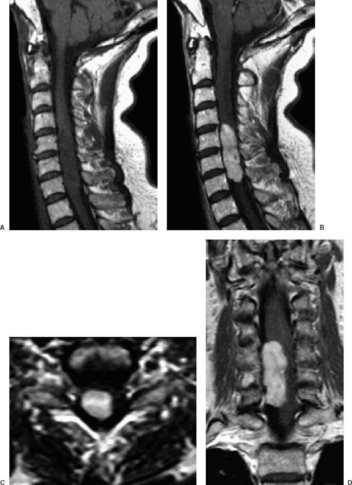
63 Diagnosis Schwannoma Problems and Tactics A very large cervical tumor significantly distorted the spinal cord of a 61-year-old woman. Diagnosis was delayed, and the patient was barely ambulatory with assistance. Urgent surgical treatment was pursued in an attempt to preserve lower extremity function. Keywords Schwannoma, cervical spine, laminoplasty A 61-year-old woman had a 3-year history of progressive right lower extremity weakness and a 2-year history of left arm numbness. She had experienced difficulty with ambulation for 6 months and required a walker. Magnetic resonance imaging (MRI) showed a 1 × 1.5 × 4.5 cm, homogeneously enhancing mass that appeared to be both intradural and extramedullary. It extended eccentrically to the right from C5 to C7 (Fig. 63–1). The patient was placed prone with slight capital flexion in the Mayfield headholder (Codman Inc., Raynham, MA). Electroencephalography (EEG) and somatosensory evoked potentials recorded before and after positioning showed no changes. A steroid bolus was administered preoperatively. A midline posterior cervical incision continued to the cervical spinous processes and lamina. A cross-table lateral radiograph was used to verify localization. When subperiosteal dissection was completed, self-retaining retractors were placed with hooks and rubber bands attached to bilateral Leyla bars® (Aesculap, San Francisco, CA) to obtain a low-profile exposure. A microangle curette was used to separate the ligamentum flavum from the superior aspect of C4 and from the inferior portion of C7 bilaterally to create an entry point for the footplate B1 bit of the Midas Rex® drill (Midas Rex Pneumatic Tools Inc., Fort Worth, TX). While the assistant pulled up on the spinous processes with a towel clamp, the laminoplasty was performed at the laminar–lateral recess junction in two motions. Drilling began at C7 and moved rostrally to C4 on one side and then was repeated contralaterally. The laminoplasty segment was set aside for later replacement. The bone edges were waxed. Two large Nu-Knit® (Johnson & Johnson, Arlington, TX) strips covered with a cottonoid patty were placed in each lateral gutter. A Spetzler Microvac sucker (Medium Malleable Stainless Suction; PMT Corp, Chanhassen, MN) was stapled to the drapes and placed into the operative field for continuous suction. The operating microscope was brought into the surgical field, and the dura was opened in the midline with a No. 15 blade. The dural incision was extended rostrally and caudally with a dural dissector. The dural leaflets were tacked up bilaterally with 4–0 Nurolon (Ethicon, Johnson & Johnson Professionals, Inc., Somerville, NJ). The dura was then dissected from the underlying arachnoid. FIGURE 63–1 (A) Preoperative sagittal T1-weighted magnetic resonance imaging (MRI) shows an apparently thickened midcervical spinal cord. (B) Sagittal, (C) axial, and (D) coronal MRIs show a large, homogeneously enhancing mass. The sagittal and axial images do not clearly indicate whether the tumor is intra- or extramedullary. The coronal image, however, clearly shows an extramedullary lesion compressing the spinal cord on the right.
Large Intradural–Extramedullary Cervical Spine Tumor
Clinical Presentation
Surgical Technique
Stay updated, free articles. Join our Telegram channel

Full access? Get Clinical Tree


